This blog has been contributed by Dr. Imran Jawaid, Consultant Ophthalmologist in Paediatric ophthalmology and strabismus surgery
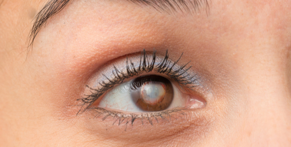
As parents, we do everything possible to support our children’s development and well-being. Vision plays a vital role in a child’s overall growth, supporting learning, movement, and communication from an early age. Yet, one area that can easily be overlooked is eyesight.
Research predicts that conditions such as myopia (short-sightedness) will increase from 27% of the world’s population to 52% by 2050. Moreover, the younger a child is when becoming myopic, the more myopic they will become and the higher the risks of vision-related complications in the future.
Let us take a closer look at why paediatric eye exams in Dubai are so crucial.
Why are regular eye exams essential for children?
Good vision is essential for a child’s development. Unlike adults, young children might not recognise or express that they have a vision problem. This is commonly the case when they have conditions such as near sightedness, farsightedness, and lazy eye. They may assume that everyone sees the way they do, which means they could struggle at school or avoid certain activities without anyone realising the actual cause. Early detection through regular eye exams for children can lead to timely interventions, allowing children to grow, learn, and fully participate in school and everyday life.
What are the most common childhood eye conditions?
Eye conditions can affect children, and early diagnosis can significantly improve treatment outcomes. Some of the most common include:
- Refractive errors: Myopia (short-sightedness), hypermetropia (long-sightedness), and astigmatism.
- Strabismus: Known as a squint or crossed eyes, which can affect depth perception and visual appearance.
- Amblyopia (Lazy Eye): This is a condition in which the brain favours one eye over the other.
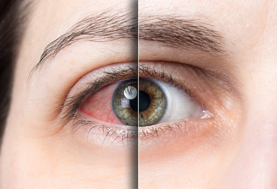
- Eye allergies and infections: These cause redness, irritation, or discharge, particularly in younger children.
When should Children have eye exams?
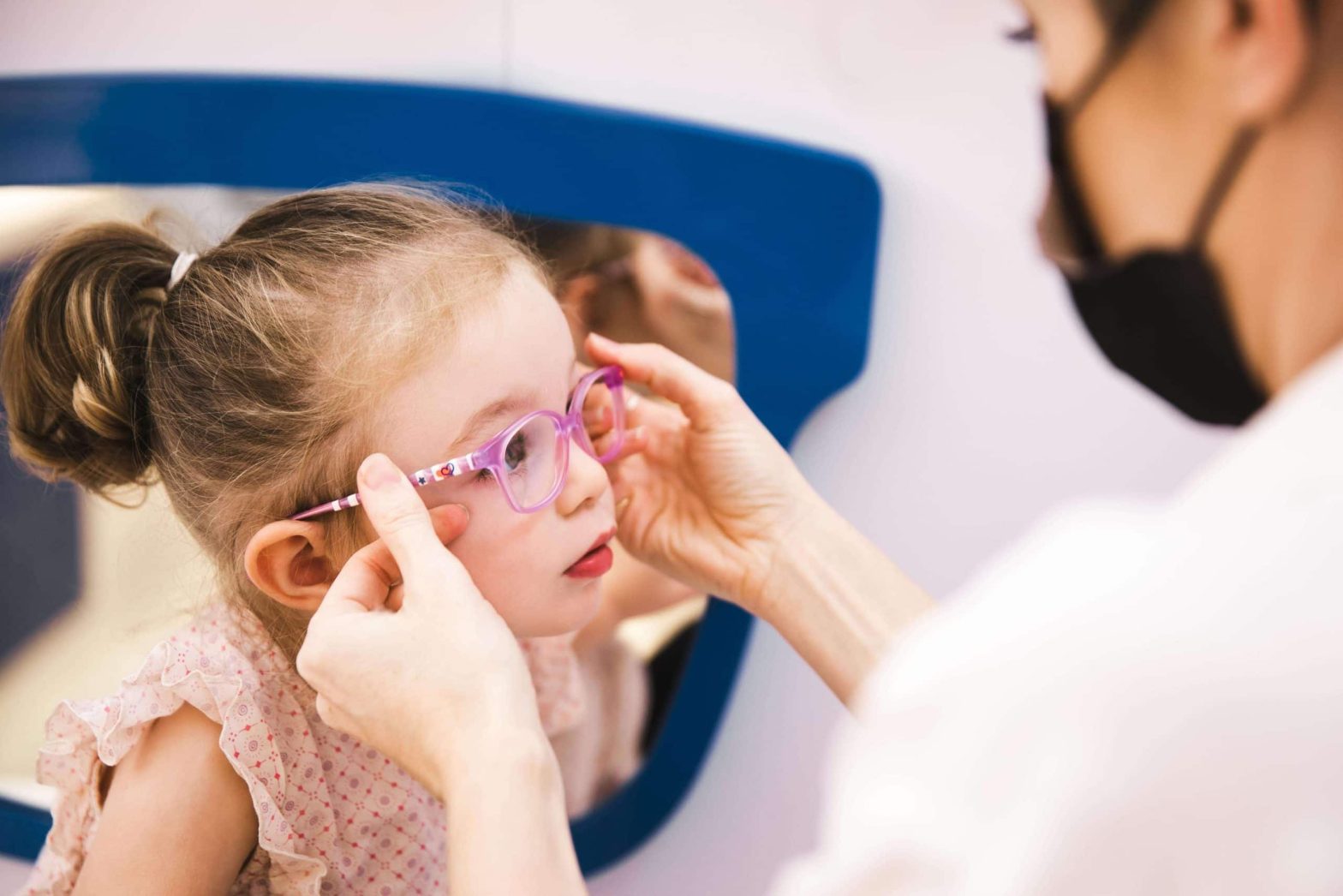
In addition to vision-related checks with your Paediatrician, we recommend that children have their first comprehensive eye exam at around six months old, followed by another at three years of age and then again before starting school. After that, exams every one to two years are advised, or more frequently if your child already has a diagnosed eye condition or significant family history.
With increased exposure to digital screens and close-up work in schools and nurseries, regular eye health monitoring is especially important. Parents should consider booking an exam with a Paediatric eye specialist if their child:
- Squints or tilts their head when looking at objects
- Sits too close to the television or holds books too close
- Struggles with reading or maintaining attention
- Complains of headaches or tired eyes
- Shows signs of clumsiness or poor coordination
- Frequently rubs their eyes or blinks excessively
What happens during a children’s vision screening?
A paediatric eye exam is tailored to suit a child’s age and developmental level. Let us take a closer look at what happens during an exam:
- Meeting Multiple Specialists: Your child will be seen by several specialists, including orthoptists, optometrists, and paediatric ophthalmologists, ensuring a comprehensive review of their eye health.
- Vision testing: Age-appropriate methods to assess how well your child sees with our highly qualified orthoptic team.
- Eye alignment and movement: This exam includes checks to see how well both eyes work together and detect any strabismus (misalignment of the eyes).
- Eye examination: The eyes’ internal structures are examined in detail, sometimes with pupil dilation or using specialist child-friendly imaging if necessary.
What are the treatment options for Children’s eye conditions
When it comes to treating your child’s eye condition, there are both non-surgical and surgical options available to help ensure they receive the best care possible. At Moorfields Eye Hospital Dubai, we provide advanced treatments supported by the latest technology:
Non-Surgical Treatments
- Eyeglasses or contact lenses: The simplest and most common treatments for refractive errors such as myopia, hypermetropia, and astigmatism in children. Eyeglasses are often the first line of treatment to help your child see better, while contact lenses offer an alternative to glasses for active children who might find glasses cumbersome.
- Myopia management includes specialist spectacle lenses, orthokeratology (Ortho-K), eyedrops (low-concentration atropine), and soft contact lenses. We will assess your child’s eyes and work with you to find the most appropriate solution to help slow down their myopia (short-sightedness). There is a wide range of options, and at Moorfields, we have access to the latest technology.
- Patching: Used primarily to treat amblyopia (lazy eye), patching involves covering the stronger eye so the weaker eye is forced to work harder. This method can help improve vision in the affected eye over time.
- Atropine occlusion: Drops can be used in the treatment of lazy eye to blur the vision in the ‘good eye’ to encourage the brain to use the weaker eye, helping to improve the vision.
Surgical Treatments
For conditions such as squint (Strabismus), where non-surgical methods did not treat the condition, surgical intervention may be recommended. This procedure corrects misalignments in the eyes, improving not only eye alignment but also depth perception and overall vision.
Final thoughts
Children’s eye health is crucial to their overall well-being. In a vibrant city like Dubai, where academic and visual demands are high, regular eye exams with children’s eye doctor are one of the most effective ways to support your child’s learning, behaviour, and social development.
If your child has not had an eye exam recently, or if you have noticed any signs of visual difficulty, it is important to get their eyes checked with a paediatric ophthalmologist. Early detection prevents future complications and gives children the best possible start in life.
At Moorfields Eye Hospital Dubai, we are here to help your child see the world clearly—now and for years to come. Let us work together to ensure your child’s vision sets them up for success.
Frequently Asked Questions
1. Can screen time affect my child’s vision? Yes, it can. With digital screens becoming a regular part of daily routines, whether for learning or leisure, there has been a noticeable rise in eye strain and early-onset myopia in children (short-sightedness). Staring at screens for prolonged periods, especially up close, can cause discomfort, blurry vision, headaches, and fatigue. It may also encourage poor visual habits, such as holding devices too close to the eyes or forgetting to blink, which affects tear film stability and eye moisture.
To help your child, include techniques like the 20-20-20 rule (taking a 20-second break every 20 minutes to look at something 20 feet away), setting appropriate screen limits, and ensuring balanced visual activities, such as outdoor playtime. These small habits can go a long way in protecting your child’s developing vision.
2. My child is afraid of doctors—how do you handle nervous children? We understand that visiting a hospital can be daunting for little ones, especially if it is their first eye exam. That is why our entire paediatric eye care environment is designed to feel calm, safe, and welcoming. Our staff are specially trained in working with children and use gentle, playful, and engaging techniques to build trust.
We allow children to familiarise themselves with the equipment, explain each step in a fun and friendly way, and offer positive reinforcement throughout. Parents are always encouraged to stay by their child’s side during the exam, and we never rush the process. Our goal is not just to complete the assessment but to make it a positive experience your child will be comfortable returning to.
3. How long does a children’s eye exam take? A typical comprehensive children’s eye exam takes around 30 to 60 minutes. The exact duration depends on your child’s age, cooperation, and whether additional tests, such as pupil dilation or retinal imaging, are needed. We always aim for a thorough yet efficient experience, balancing accuracy with your child’s comfort.
You will have time to discuss any concerns, and we will ensure your child gets the focused attention they deserve. We recommend allowing a little extra time in your schedule, especially for first-time visits or if your child has specific vision needs.
4. Can poor vision affect my child’s behaviour or school performance? Yes, and quite significantly. Many children with vision issues may appear distracted, inattentive, or reluctant to engage in activities like reading or sports, not because of behavioural problems but simply because they are not seeing clearly. This can lead to frustration, low self-esteem, or even being misdiagnosed with learning difficulties.
A simple eye exam can reveal if vision is the underlying cause. With early intervention, whether through glasses, eye exercises, or other treatments, we can make a dramatic difference in your child’s ability to learn and thrive both academically and socially.
5. Do you offer eye care for children with special needs? Absolutely. We are proud to offer inclusive and compassionate eye care for children with developmental delays, autism, Down syndrome, and other neurological, behavioural or learning conditions. Our team takes extra time and care to understand each child’s unique needs, tailoring our approach accordingly.
Whether it means adapting communication, simplifying tests, or creating a sensory-friendly environment, we aim to make every child’s experience as smooth and positive as possible. Every child deserves clear vision; we are here to help them achieve it.
6. How often should children with glasses or existing conditions be seen? Children who already wear glasses or have known vision conditions should typically be seen every 6 to 12 months, depending on their case. Frequent monitoring allows us to check for prescription changes, ensure treatments are working effectively, and make timely adjustments.
At Moorfields Eye Hospital Dubai, we offer ongoing care and support as your child grows. Regular follow-ups are a key part of safeguarding their long-term visual development.
This blog on Oculoplastic Surgery has been contributed by Dr. Rafic Antonios, Specialist Oculoplastic Surgeon.
Oculoplastic surgery, often known as eyelid surgery, is a specialised field of medicine focusing on the areas around the eyes and the face. This includes the eyelids, orbit (eye socket), and lacrimal (tear) system. The purpose of these procedures ranges from aesthetic enhancements to significant functional improvements, including improvements in vision.
The Interplay Between Oculoplastic Surgery and Vision
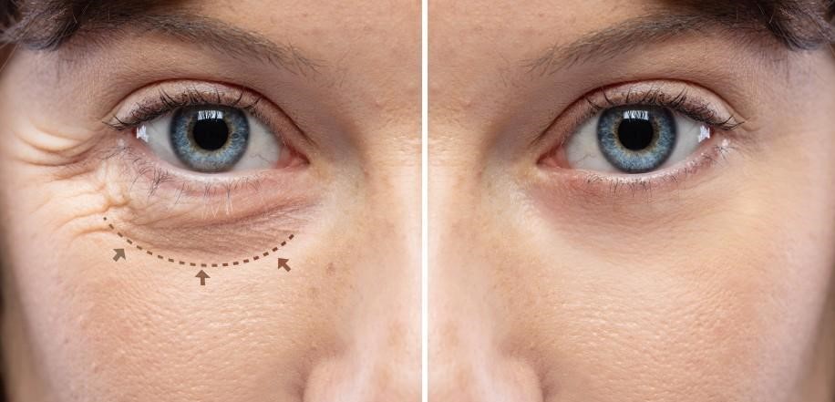
Generally, the eyes and the surrounding structures work harmoniously to deliver clear, uninterrupted sight. However, droopy eyelids (ptosis) or eyelid malposition can disrupt this equilibrium, impairing vision. That’s where oculoplastic surgery steps in.
Addressing Common Concerns Through Surgery
Droopy eyelids, a common age-related condition, can obstruct the field of vision, making daily activities like reading or driving challenging. Oculoplastic surgery corrects this by tightening the muscles that lift the eyelid, thereby widening the visual field. Similarly, conditions like entropion (inward-turning eyelid) or ectropion (outward-turning eyelid) can cause discomfort, tear overflow, and blurred vision. Surgery to reposition the eyelid reinstates normalcy, relieving the symptoms and restoring vision clarity.
The Transformative Potential of Oculoplastic Surgery
In conclusion, oculoplastic surgery is not just about cosmetic enhancement. It’s a field aimed at restoring or maintaining the eye’s optimal functionality, significantly impacting vision. While the thought of surgery can seem daunting, understanding its potential benefits can provide reassurance. Consulting with an oculoplastic surgeon could be a transformative step for your vision if you are experiencing vision disruption due to eyelid or surrounding orbital structure issues.
If you are considering oculoplastic surgery in Dubai or want to learn more about how these procedures can enhance your vision and appearance, we are ready to help you by providing personalised care tailored to your unique needs and goals.
Orthoptist Sehar Mann has contributed to this blog on Stages of visual development in Children.
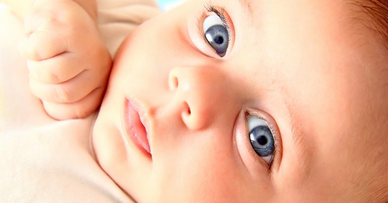
We are all born with blurry vision, and our vision develops in the first 7-8 years of life but is most important when we are babies and toddlers.
Understanding the stages of visual development in babies and toddlers and how they perceive colours can help you provide the correct visual stimulation in their early months of life.
Stages of Visual Development
- The Newborn Stage (0-2 months): Newborn babies can only see objects about 8 to 12 inches from their face – roughly the distance between your face and theirs during feeding or cuddling. They are drawn to high-contrast patterns, like black and white since their eyes are still developing.
- The Infancy Stage (2-4 months): During this period, your baby’s vision starts to improve gradually. They follow moving objects and show interest in colourful toys and pictures. It is normal for their eyes to cross or wander as their muscles strengthen occasionally.
- The Exploratory stage (4-6 months): At 4 to 6 months, babies reach for objects and have better depth perception as they can see more colours more clearly. This is an ideal time to introduce more colourful toys and books with large, bold pictures to stimulate their developing vision.
How Toddlers Perceive Colours
Understanding how toddlers perceive colours can help you create an engaging and stimulating environment for them. Here are some tips to help them:
- High Contrast is Key: In the early months, your baby is drawn to high-contrast colours, such as black and white because they are easier for their developing eyes to distinguish. Consider incorporating these colours into your baby’s nursery decorations or toys.
- The world of Pastels: As babies grow, they will notice more subtle colours like pastels. Soft blues, pinks, and yellows can be soothing and visually appealing for them.
- Vibrant Colours: Babies can see a broader range of colours in the toddler stage. Bright and vibrant colours like red, green, and blue can capture their attention and stimulate their visual development.
Visual Stimulation in the First 6 Months
Visual stimulation is essential for development during the first six months. Here are some simple ways to provide the correct visual experiences:
- Face-to-Face Interaction: Spending quality time making eye contact and talking to your baby can help develop their vision. They love to gaze at your face, and this helps strengthen their visual focus.
- Colourful mobile Toys: Hang a colourful mobile above the crib. The gentle movement and bright colours will intrigue your baby and encourage them to track the objects.
- Picture Books: Choose picture books with large, bold images and vibrant colours. Reading to your baby fosters bonding and enhances their visual recognition skills.
- Tummy Time: Give your baby plenty of tummy time. This helps them develop strong neck and shoulder muscles, which are essential for visual exploration.
- Mirrors: Babies are often fascinated by their reflection. A baby-safe mirror can provide endless entertainment and help with self-recognition.
Watching your toddler’s visual development is an exciting journey that requires your support and engagement. By understanding the stages of visual development and how your baby perceives colours, you can create a stimulating environment that encourages healthy growth and curiosity. Remember, every baby is unique, so enjoy the discovery process together and celebrate each new milestone.
At Moorfields Eye Hospital Dubai, our dedicated paediatric department is staffed with expert ophthalmologists, optometrists, and orthoptists, all passionately dedicated to the eye care needs of children.

Strabismus, commonly called ‘crossed eyes,’ impacts approximately 2% – 4% of children worldwide. This condition results in the misalignment of the eyes, causing them to point in different directions and disrupting binocular vision.
Strabismus is primarily seen in infants and young children and is not just a cosmetic concern. It can significantly impact vision development and depth perception, essential for numerous activities, from catching a ball to crossing a street. When the eyes do not align properly, the brain may start to ignore the image from the eye that is not aligned to prevent seeing double, causing ‘amblyopia,’ also known as a lazy eye.
Emphasising early detection is crucial with strabismus, as the initial years of a child’s life are vital for visual development. Any signs of the condition, such as squinting, tilting the head while looking at objects, or noticeable misalignment of the eyes, should prompt an immediate medical consultation.
The treatment journey for a child with strabismus often involves a team of specialists. An optometrist performs regular eye exams and prescribes glasses if necessary. An orthoptist specialising in strabismus and amblyopia devises personalised eye exercises to improve eye coordination. Lastly, a paediatric eye consultant oversees the child’s treatment, deciding when surgical intervention is necessary and ensuring the various treatments align to maximise visual development and overall quality of life.
In essence, strabismus affects not only the physical appearance but also the functional vision of a child. Therefore, recognising and addressing strabismus in its early stages is crucial for optimal eye health and developmental growth.
If you notice any of the signs or symptoms of Strabismus, please seek care from a specialised ophthalmologist to help your child’s vision develop correctly.
At Moorfields Eye Hospital Dubai, our dedicated paediatric department is staffed with expert ophthalmologists, optometrists, and orthoptists, all passionately dedicated to the eye care needs of children.
This blog on Selective Laser Trabeculoplasty for glaucoma treatment was authored by Dr. Salman Waqar, a Consultant Ophthalmologist in Cataract and Glaucoma Surgery.
Living with glaucoma often means meticulous eye drop management and close intraocular pressure (IOP) monitoring. Thankfully, advancements in eye care, such as Selective Laser Trabeculoplasty (SLT), offer a more refined to manage this condition.
What is SLT?
Selective Laser Trabeculoplasty is a minimally invasive procedure designed to help control glaucoma by decreasing IOP. One of its standout features is the potential to lessen, or in some cases, even eliminate, the dependence on glaucoma eye drops, especially beneficial for those with open-angle glaucoma.
How Does It Work?
Imagine the eye’s drainage system, the trabecular meshwork, as a sink’s drain. Over time, this drain can become less efficient, leading to increased IOP. During this procedure, a precision-targeted laser is applied to this drainage system. Unlike other methods, Selective Laser Trabeculoplasty ensures no thermal damage to the nearby tissue. The treatment encourages the drainage system to work more efficiently by promoting beneficial biological changes within its cells. As the drainage improves, the IOP naturally decreases.
Why Consider SLT?
Selective Laser Trabeculoplasty is revolutionary in its approach. Unlike more aggressive surgeries, it targets only specific cells, ensuring surrounding tissues remain untouched. This selectivity means a less invasive procedure, minimal risks, and quicker recovery times.
However, it’s essential to remember that while it is highly effective, it might not completely negate the need for glaucoma eye drops for all patients. For some, it can notably decrease the amount or frequency of medication, leading to improved comfort and better adherence to treatment.
In Conclusion
Selective Laser Trabeculoplasty is a promising option in glaucoma care. Potentially reducing our reliance on daily eye drops offers a more convenient yet effective way to keep glaucoma in check and safeguard our vision. If you are considering this procedure, discussing it with a glaucoma specialist is recommended to tailor the best care for your eyes.
This blog has been contributed by Dr. Luisa Sastre, Consultant Ophthalmologist in Medical retina and cataract surgery.
Cataracts, a common eye condition characterised by clouding of the lens, can affect individuals of all ages. However, people with diabetes are particularly susceptible to developing this condition. In this blog, we will explore the connection between them, understand the underlying mechanisms, and discuss the treatment of cataracts in diabetics.
The connection: Diabetes, a metabolic disorder affecting blood sugar regulation, increases the risk of complications, including eye-related issues such as diabetic retinopathy. Cataracts occur when proteins in the lens clump together, causing clouding and reduced vision. People with diabetes are more likely to develop cataracts at an earlier age and experience faster progression than those without diabetes.
Mechanisms Behind Cataract Formation in Diabetes:
Diabetes can cause cataracts by increasing sugar levels in the body. This can lead to imbalances and changes in the lens structure, making it less transparent. Diabetes can also cause damage to lens proteins, which makes cataracts form faster.
Managing the Risks of Cataracts in Diabetes:
Effective management of cataract risks in individuals with diabetes is crucial. Maintaining stable blood sugar levels through proper diabetes management helps slow cataract progression. In addition, regular eye examinations are essential for early cataract detection. Adopting a healthy lifestyle, including a diet rich in antioxidants, regular exercise, and avoiding smoking, can also reduce the risk of cataracts.
Cataract treatment options for people with diabetes: Surgery is the most effective treatment for cataracts. The procedure involves removing and replacing the clouded lens with an artificial intraocular lens (IOL). People with diabetes must work closely with their doctors to ensure stable blood sugar levels before, during, and after surgery, minimising the risk of complications and optimising visual outcomes.
Understanding the connection between those two conditions and managing the associated risks is crucial for maintaining good eye health. Individuals can effectively navigate this coexisting condition by controlling blood sugar levels, regular eye check-ups, and considering cataract surgery when necessary.

This blog has been contributed by Dr. Ammar Safar, Consultant Ophthalmologist and Vitreoretinal Surgeon and Chief Medical Officer.
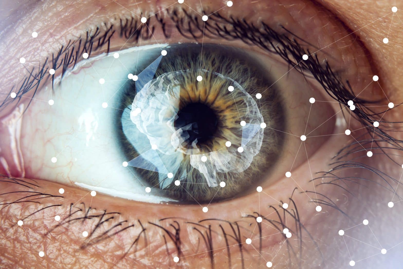
Artificial intelligence (AI) has revolutionised various industries, and it’s making its way into ophthalmology. AI technologies are being viewed as valuable tools for diagnosing, managing, and treating eye diseases.
AI algorithms can analyse large amounts of data in a short time, making it easier to identify and treat eye conditions. One of the most significant applications of AI in ophthalmology is in detecting and diagnosing retinal diseases, including diabetic retinopathy, age-related macular degeneration, and glaucoma.
Diabetic retinopathy is a leading cause of blindness in adults. It is unfortunately very prevalent in our part of the world with a large number of patients affected. However, with the help of AI, ophthalmologists can now detect the disease early and provide timely treatment. AI-powered retinal imaging systems can analyse a large number of retinal images for early signs of the disease with an outstanding level of accuracy, providing ophthalmologists with a more accurate diagnosis and making it next to impossible to delay or miss a diagnosis.
Age-related macular degeneration is another eye condition that can lead to vision loss. AI algorithms can analyse the retina’s images to detect signs of the disease and track its progression, enabling ophthalmologists to provide appropriate treatment.
Glaucoma is a condition that damages the optic nerve, leading to vision loss. AI technologies can detect the disease in its early stages, allowing for early intervention to prevent further vision loss. AI algorithms can analyse visual field tests, imaging tests, and other data and compare them to previous tests detecting even the very minute variations providing a very powerful tool to determine the likelihood of developing glaucoma and its progression.
AI can also assist ophthalmologists in the management of cataracts, which is a common cause of vision loss in older adults. AI algorithms can analyse patient data, including medical history, to determine the appropriate surgical approach and intraocular lens power.
AI technology has the potential to revolutionise ophthalmology, enabling ophthalmologists to provide more accurate diagnoses and personalised treatment plans. While AI cannot replace human expertise, it can significantly enhance it, providing ophthalmologists with a powerful tool to improve patient care.
In conclusion, AI can transform ophthalmology by improving the diagnosis, management, and treatment of eye diseases. As technology advances, AI will undoubtedly become an increasingly essential tool for ophthalmologists.
This blog has been contributed by Dr. Salma Yassine, Consultant Ophthalmologist in Paediatric & Neuro-ophthalmology
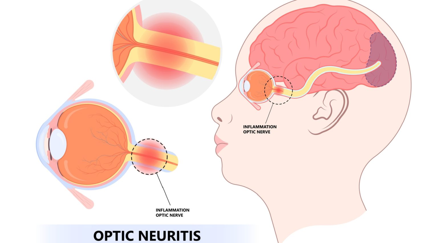
What is optic neuritis?
Optic neuritis occurs when our immune system mistakenly targets the substance covering your optic nerve, resulting in inflammation and damage to the myelin. This disrupts electrical impulses that travel from the eye to the brain, causing blurred or dark vision.
How does optic neuritis affect me?
Optic neuritis usually affects one eye. Symptoms might include:
- Pain: Eye pain that’s usually worsened by eye movement. Sometimes it presents as a dull ache behind the eye.
- Vision loss in one eye: Noticeable vision loss usually develops over hours or days and improves over several weeks to months. However, vision loss can be permanent in some people.
- Visual field loss: Central or peripheral vision loss
- Loss of colour: Colours appear less vivid
What causes optic neuritis?
The following autoimmune conditions often are associated with optic neuritis:
- Multiple sclerosis: It is a disease in which the autoimmune system attacks the myelin sheath covering nerve fibres in your brain. The risk of developing multiple sclerosis after optic neuritis increases further if an MRI scan shows demyelinating brain lesions
- Neuromyelitis Optica: In this condition, the inflammation affects the optic nerve and spinal cord. As a result, it often results in diminished visual recovery after an attack compared with MS.
- Myelin oligodendrocyte glycoprotein (MOG) antibody disorder: Like neuromyelitis optica, recurrent attacks of inflammation can occur in the optic nerve, spinal cord or brain. However, recovery from MOG attacks is usually better than recovery from neuromyelitis optica.
When symptoms of optic neuritis are more complex, other associated causes need to be considered, including infections, rheumatological diseases, and drugs or toxins (ethambutol or methanol)
Why do I need to see a neuro-ophthalmologist?
- Neuro-ophthalmologist is experienced in sorting out the differences between optic neuritis and other optic nerve diseases.
- During your office visit, the doctor will check your visual fields and scan your optic nerves
- Your doctor will order an MRI of the brain with special views of the orbits with contrast to confirm optic neuritis
- Your doctor may order other tests, such as blood tests or a chest X-ray
What are the possible complications?
- Optic nerve damage: Most have permanent optic nerve damage after an episode of optic neuritis, but the damage might not cause permanent symptoms.
- Decreased visual acuity: Most people regain normal or near-normal vision within months, but a partial loss of colour discrimination might persist.
Side effects of treatment: Steroid medications used to treat optic neuritis suppress the immune system, which causes your body to become more susceptible to infections. It can also cause mood changes and weight gain.
This blog has been contributed Dr. Salman Waqar, Consultant Ophthalmologist in Cataract and Glaucoma Surgery.
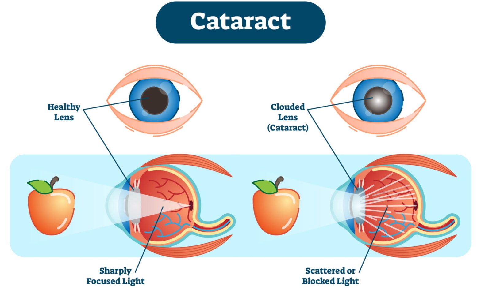
Cataracts are a common age-related eye condition where the eye’s lens becomes cloudy and opaque, affecting vision. The lens is a clear, elastic structure that helps focus light onto the retina, which sends visual signals to the brain. As we age, the lens becomes less flexible and loses its transparency, leading to cataracts.
There are several types of cataracts, including congenital cataracts (present at birth), traumatic cataracts (caused by injury), and secondary cataracts (caused by other eye conditions or diseases). However, the most common type is age-related cataracts, which occur naturally with age.
Cataracts can cause various symptoms, including blurred or hazy vision, glare or halos around lights, double vision in one eye, frequent changes in eyeglass or contact lens prescriptions, fading or yellowing of colours, and difficulty seeing at night. If left untreated, cataracts can eventually lead to vision loss.
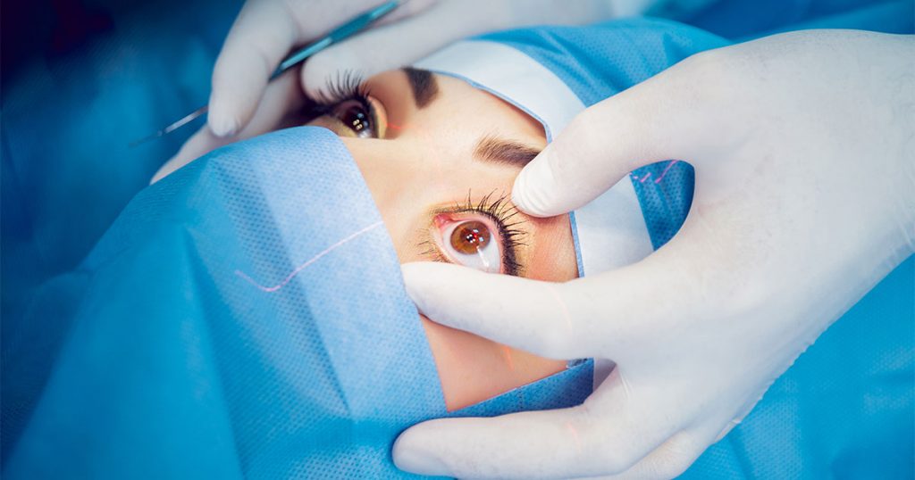
Cataracts are usually managed through surgery. In cataract surgery, the cloudy lens is removed and replaced with an artificial lens called an intraocular lens (IOL). Cataract surgery is a safe and effective way to restore vision and improve quality of life.
Before deciding on cataract surgery, an eye doctor will perform a comprehensive eye exam to determine if cataracts cause the patient’s vision problems. If cataracts are diagnosed, the eye doctor will evaluate the extent of the cataract and recommend the best course of treatment.
After cataract surgery, patients typically need eye drops for a few weeks to help prevent infection and inflammation. It is also important to protect the eyes from injury, such as by wearing sunglasses outside. However, most patients experience improved vision within a few days or weeks after surgery.
In conclusion, cataracts are a common age-related eye condition that can cause vision problems. Cataract surgery is a safe and effective way to treat cataracts and restore vision. If you are experiencing cataract symptoms, you must see an eye doctor for a comprehensive eye exam and evaluation. With advances in surgical techniques and technology, cataract surgery has become a highly effective way to improve vision and quality of life.

This blog on glaucoma awarness month has been contributed Dr. Salman Waqar, Consultant Ophthalmologist in Cataract and Glaucoma Surgery.
Glaucoma occurs when there is high pressure in the eye, which can lead to reduction or loss of vision due to damage to the optic nerve (the nerve that transmits signals from our eye to the brain). It is one of the leading causes of blindness worldwide, with nearly 60 million people already affected. This number is projected to increase by almost fifty percent over the next ten years.
January is Glaucoma Awareness Month and is an opportunity for us all worldwide to highlight this blinding condition (also known as the “silent thief of sight”).
Early detection and prompt treatment are essential. The campaign aims to increase awareness of the importance of annual eye examinations to diagnose and treat the condition early before vision is harmed for our loved ones and us.
Typically, your eye specialist will conduct the following very specialised and precise tests to screen for glaucoma:
- Eye pressure check
- Clinical evaluation of the optic nerve
- Visual Field testing
- Optical Coherence Tomography (OCT) scan of the nerves
If a diagnosis of glaucoma is made, treatment can be done with easy-to-use eye drops, gentle lasers or, in more advanced cases, with the latest surgical techniques. However, the treatment decision can vary from person to person, and your eye specialist will recommend a bespoke treatment plan keeping in mind your individual requirements.
For more information, please visit www.glaucoma.org”
This blog on Neuro-ophthalmology has been contributed by Dr. Salma Yassine, Consultant Ophthalmologist in Paediatric & Neuro-ophthalmology
What is neuro-ophthalmology?
Neuro-ophthalmology is an ophthalmic subspecialty that addresses the relationship between the eye and the brain. The optic nerve acts as a cable that connects what we see through our eyes to the brain. The brain then changes the visual signals into images and helps us know what we are seeing. If the optic nerve is damaged, these visual signals are impaired, which leads to decreased visual perception. The field of neuro-ophthalmology deals with such neurological conditions that affect the eye and causes problems with vision. Some neuro-ophthalmic disorders can cause permanent damage if not diagnosed and treated adequately.
What are the most common symptoms of neuro-ophthalmic diseases?
Some common Neuro-ophthalmology symptoms include reduced vision, double vision, headaches, impaired colour vision, and visual field cuts. These symptoms are not to be ignored, if you face any of them, you must immediately seek advice from a neuro-ophthalmologist
What are common types of neuro-ophthalmic diseases?
A few of the most common neuro-ophthalmic conditions are optic neuritis, ischemic optic neuropathy, comprehensive optic neuropathy (pituitary tumours), papilledema, inflammatory and infectious optic neuropathies, cerebrovascular disorders involving vision, tumours involving vision, blepharospasm and hemifacial spasm, nystagmus, thyroid eye disease, myasthenia gravis, ocular motor disorders, pupillary abnormalities, hereditary optic neuropathies in patients who have unexplained vision loss
Some disorders that require immediate attention from neuro-ophthalmologists include;-
- Optic neuritis: It is an eye disorder that is caused by inflammation of the optic nerves. This inflammation can be caused by infection or autoimmune disorders like multiple sclerosis and neuromyelitis optica. It is a disease that usually affects young adults in 1 eye but often affects 2 in children. Patients with optic neuritis can develop nagging eye pain, pain with eye movements, blurry vision, and loss of colour perception. These issues can be serious because it can lead to permanent vision loss.
- Papilledema : It is a condition in which optic nerves swell up due to increased intracranial pressure. It is often accompanied by headaches, dimming of vision and rushing noises in the ears. Papilledema can lead to optic atrophy and blindness if not treated in a timely manner. Sometimes papilledema can be a warning sign for a tumour or haemorrhage. If papilledema is not traced to a particular problem, then it is called idiopathic cranial hypertension.
- Nutritional optic neuropathy: Toxic substances found in alcohol and tobacco can also damage the optic nerve. Certain vitamin deficiencies like folic acid and vitamin B complex can cause optic neuropathy
- None ischemic arthritic optic neuropathy (NAION): There are many risk factors for NAION, some of which include uncontrolled sugar levels in diabetics or high blood pressure in hypertensive patients. These systemic conditions can affect the blood vessels that supply the optic nerve, which can result in disabling vision loss.
- Strabismus (squints): It is a disorder when both eyes cannot align in one direction, which can cause double vision. Paralytic strabismus occurs when the muscles are unable to move the eye, disrupting the coordination between both eyes. This could be due to a lesion compressing the nerves that connect to the ocular muscles or due to microvascular damage to these nerves
This blog on Dry eyes has been contributed by Dr. Alia Issa, General Ophthalmologist at Moorfields Eye Hospital Dubai
Introduction
Dry eye is one of the most common reasons to visit an ophthalmologist. It is estimated that around 75% of people worldwide suffer from dry eye at some point in their life.
What causes dry eye?
Environmental factors like air conditioning, air pollution, cigarette smoke, contact lens use, dusty and hot weather, and extended computer and smartphone use can contribute to dry eye and ultimately worsen symptoms. But also gender and age, systemic diseases, especially autoimmune diseases and medications play an essential role in developing dry eye disease.
What are the symptoms?
Symptoms can include foreign body sensation, pain, itchiness, tearing, redness of the eyes and blurry vision.
How do we diagnose dry eye?
When diagnosing a dry eye we differentiate between two main types of dry eye i.e. evaporative dry eye and aqueous deficient dry eye. Evaporative dry eye means that the tear film evaporates from the ocular surface faster than it should. In contrast, aqueous deficient dry eye describes a condition where tear production is reduced. Both have different underlying causes, which require different treatment approaches.
How do we treat dry eye?
Dry Eye is a complex disease; very often, it is not one but multiple factors that contribute to it. Therefore, a detailed history, slit lamp examination and special diagnostic tests are vital in finding the right individual treatment approach for each patient. Treatment options can vary from lifestyle changes, different types of artificial tears, and tear plugs to in-office treatments like Intense Pulse Light (IPL) , depending on the underlying causes of the dry eye.
At Moorfields Eye Hospital Dubai, we can tailor an individual treatment plan for each patient. If you feel you are experiencing dry eye symptoms, we recommend you visit a specialist to get a professional diagnosis and treatment if required.