
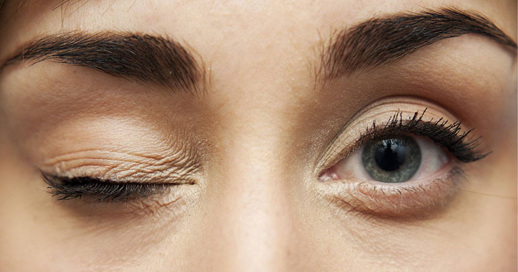
This week’s blog on Reasons Why You Shouldn’t Rub Your Eyes has been contributed by Dr Luisa Sastre, Specialist Ophthalmologist in Medical Retina
5 Reasons Why Rubbing/Touching Your Eyes Is Not A Good Idea
Many of us tend to rub or touch our eyes frequently (consciously or not) during the course of every day. But did you know that every time we do this, we are taking serious risks?
Here is a list of eye diseases that have a connection with touching or rubbing your eyes:
- INFECTIOUS CONJUNCTIVITIS – Adenoviral conjunctivitis is the most common cause of ‘red eye’ in the world and is very easily transmitted by someone who is infected. The virus moves around through direct contact with eye and nasal secretions or by indirect contact with contaminated surfaces (door handles, elevator buttons…). You can even get the virus from someone who has no symptoms but is incubating the disease, days before the symptoms appear. This is one of the reasons why touching your eyes is not a good idea.
- KERATOCONUS – Keratoconus is an eye condition which results in a deformed and progressive thinning of the cornea, at the front of the eye. Keratoconus can cause severe loss of vision and surgery might be needed to stop it progressing. Rubbing your eyes is a known risk factor in the development of keratoconus and can make the condition worse.
- ALLERGIC CONJUNCTIVITIS – Allergic / atopic conjunctivitis is caused by allergens (irritants such as dust, pollen, pollution) in the environment or in the foods we eat. It causes itching which can sometimes be severe and our natural reaction is to rub our eyes. However, this rubbing action also stimulates the release of histamine, which causes even more itching and the desire to keep rubbing. The process can result in even more allergens and irritants, including bacteria, fungus and viruses, getting into to the eye.
- CORNEAL ULCERS – Dry eye syndrome is uncomfortable and feels like you have something in your eye and leads to rubbing. Rubbing can help produce tears that will soothe the irritation temporarily. However if left untreated, dry eye syndrome can eventually cause small wounds in the surface of your eye (keratitis) and rubbing can turn a mild keratitis into a corneal ulcer. Furthermore, rubbing your eye to remove something entered into it, can cause corneal ulcers and worsening of the condition (e.g. small metal splinters from grinding or cutting machines lodged on the cornea or conjunctiva are one of the most common emergencies seen by ophthalmologists). Finally, ‘fighting with a contact lens that won’t come out’ or that you think is ‘lost in your eye’, can cause significant irritation and corneal ulcers.
- AFTER SURGERY – No matter how much you would like to rub your eyes, rubbing them after eye surgery is extremely dangerous. Rubbing your eyes can open surgical incisions causing leakage and transmission of bacteria and viruses, weeks or months after surgery. It can also move or dislodge a flap after LASIK surgery or an intraocular lens.
What are the alternatives to rubbing?
No matter how much pleasure and relief you find in rubbing, compressing or touching your eyes, it can be dangerous.
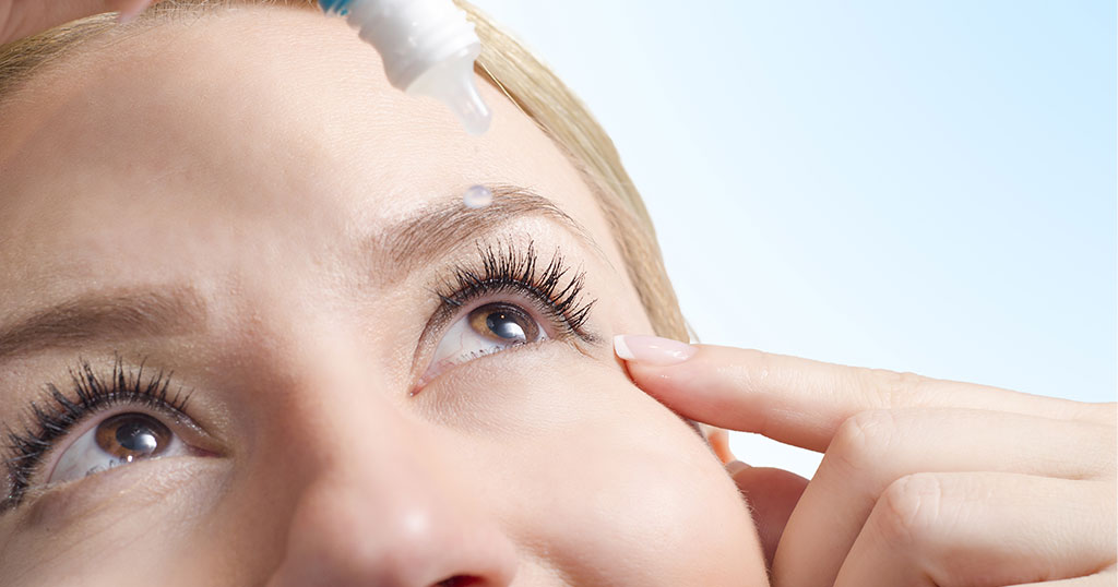
Although we know that rubbing the eyes creates tears that moisturise the eyes, it is always much safer to use artificial teardrops, whenever you have the desire to rub. There is no limit to the use of artificial teardrops and you can use them hourly or even every 15- 30 minutes, if you have significant discomfort. Preservative-free artificial teardrops are the best option. And of course, visit your ophthalmologist if the itchiness or the need to rub continues, as it can be a symptom of a condition that might need treatment.

Good vision plays an important role in a child’s education, development, and confidence. Seeing clearly is not just about reading a blackboard or a book; it influences how children interact with the world, communicate with others, and absorb information.
Research suggests that up to 80% of learning in a classroom is visual, meaning that undetected vision problems can significantly affect a child’s ability to learn and participate fully at school.
For parents, teachers, and caregivers, recognising potential signs of a vision concern early can make an important difference. Seeking a timely assessment with a paediatric eye doctor helps ensure that children receive the appropriate support for their visual development, learning, and everyday activities.
What Are the Signs That May Indicate Vision Problems in Children?
Children do not always recognise or explain when something is affecting their vision. Instead, they may display behaviours that suggest they are having difficulty seeing clearly. Common signs include:
- Holding books, tablets, or phones very close to the face
- Squinting, tilting the head, or covering one eye to focus more clearly
- Complaining of blurred or double vision
- Frequent eye rubbing or excessive blinking
- One eye appears to turn inward or outward
- Headaches, particularly after reading, studying, or screen use
- Difficulty reading, writing, or copying from a board
- Avoiding reading or other close-up tasks
- Struggling to maintain attention during visually demanding activities
If any of these signs appear regularly, a comprehensive eye examination with a paediatric eye specialist is recommended. These signs do not always indicate a serious condition, but a professional assessment ensures that any underlying concern is identified and managed at the earliest opportunity.
What Eye Conditions Affect Children’s Vision?
A number of eye conditions can affect how clearly a child sees. Some of the most common conditions include:
- Short-sightedness (myopia): A condition where distant objects appear blurred.
- Long-sightedness (hyperopia): A condition where close-up objects may appear blurred.
- Astigmatism: An irregularity in the shape of the cornea or lens that causes vision to appear blurred or distorted at all distances.
- Amblyopia (lazy eye): A condition where one eye does not develop normal vision, often because the brain begins to favour the stronger eye.
- Strabismus (Squint): A condition in which the eyes are not properly aligned, causing one eye to turn inward, outward, upward, or downward.
What Are the Vision-Related Learning Challenges for a Child?
When a child has an undetected vision problem, it can affect their classroom experience and their ability to fully engage in learning activities. In some cases, children may experience challenges such as:
- Spoken language: Delayed speech development or difficulty following verbal instructions.
- Written language: Challenges with reading, writing, spelling, or understanding text.
- Working with numbers: Difficulty tracking numbers, copying from the board, or aligning columns of figures.
These learning difficulties may sometimes be mistakenly attributed to attention problems or behavioural issues, when in fact a visual concern may be contributing. Early identification and treatment by a Paediatric Ophthalmologist can help children stay engaged and confident in their learning.
Why Does Early Detection of Children’s Eye Conditions Matter?
The early years of a child’s life play an important role in the development of vision. During childhood, particularly between the ages of two and seven, the visual pathways between the eyes and the brain are still developing. If certain vision problems are not identified during this period, the brain may not learn to process visual information as effectively as it should. In some cases, this can lead to lasting visual difficulties, even if the eyes themselves are structurally healthy.
When issues are present from birth, children may not always realise that their vision is affected, as that is simply what they have grown accustomed to. This is why routine eye examinations and attentive observation from parents, teachers, and caregivers are so important. The earlier a concern is identified, the greater the opportunity for effective management and support.
Can Vision Problems in Children Be Corrected?
The good news is that many childhood vision problems can be effectively managed, especially when identified early. The type of treatment depends on the specific condition and its severity, but common approaches include:
- Prescription glasses or contact lenses to correct refractive errors such as short-sightedness, long-sightedness, or astigmatism
- Patching therapy, where the stronger eye is covered for a set period each day to encourage the weaker eye to develop, is commonly used in the management of amblyopia
- Specialist exercises or orthoptic therapy to improve how the eyes work together, particularly in cases of eye alignment issues
- Ongoing monitoring by a paediatric eye specialist to track visual development and adjust treatment as needed
With the right support, many children experience significant improvements in their vision, which in turn can positively affect their confidence, learning, and overall quality of life.
Advice to support Children’s vision in Dubai?
In cities such as Dubai, children are often introduced to digital devices, including tablets, computers, and smartphones, at an early age. Increased time spent on near activities, such as reading or screen use, combined with reduced time outdoors, has been associated with a higher likelihood of developing short-sightedness. In addition, global research suggests that myopia is increasing worldwide, with projections estimating that around half of the world’s population may be affected by 2050.
There are several practical steps that parents and caregivers can take to help support healthy visual habits during childhood:
- Follow the 20-20-20 rule: After every 20 minutes of screen use or close-up work, encourage your child to look at something approximately 20 feet away for at least 20 seconds. This helps reduce eye strain during prolonged near-focus activities.
- Encourage regular outdoor time: Spending time outdoors, ideally around one to two hours each day, has been linked to a reduced risk of developing short-sightedness in children. Natural daylight plays an important role in healthy visual development.
- Set healthy screen boundaries: Establish age-appropriate limits on daily screen time and encourage regular breaks during extended use.
- Maintain a healthy viewing distance: Encourage children to hold books, tablets, and other devices at a comfortable arm’s length rather than very close to their faces.
- Schedule regular eye examinations: Routine checks with a paediatric eye specialist can help identify any emerging concerns early, even if no symptoms are present.
When to Consider an Eye Examination for Children?
You should consider a professional assessment if:
- You observe several of the vision-related signs noted above
- Your child is finding reading, writing, or other visually demanding tasks challenging
- There is a family history of eye conditions or vision problems
- Your child frequently complains about their vision or seems uncomfortable during activities that require focusing
Early eye examinations are a simple and proactive way to support your child’s overall health and development. A paediatric eye specialist can accurately diagnose underlying concerns and recommend the most appropriate treatment or visual correction.
Paediatric Eye Care at Moorfields Eye Hospital Dubai
At Moorfields Eye Hospital Dubai, a dedicated paediatric department supports children’s eye health through every stage of development. The team brings together ophthalmologists, orthoptists, and optometrists who collaborate to provide a thorough, holistic assessment of each child’s vision.
This multidisciplinary approach means that during a single visit, your child’s eyes can be assessed from multiple perspectives, helping to identify any concerns that may require attention. From initial screening through ongoing management, the team is experienced in working with children of all ages in a calm, reassuring environment, ensuring that both children and their families feel supported throughout the process.
Conclusion
Healthy vision plays an important role in a child’s ability to learn, explore their surroundings, and participate confidently in everyday activities. Because children may not always recognise or communicate when their vision is affected, early signs can sometimes be overlooked. Supporting good visual habits, encouraging time spent outdoors, and seeking professional advice when concerns arise can all help protect a child’s long-term eye health.
Furthermore, being aware of behaviours that may indicate a vision concern and ensuring children have their eyes checked when needed can help support healthy visual development. Early assessment allows eye care professionals to identify potential issues and recommend appropriate guidance or treatment where necessary.
Frequently Asked Questions (FAQs)
At what age should my child have their first eye examination?
Children should ideally have their first comprehensive eye examination before starting school, typically between the ages of 3 and 5. At this stage, vision and visual coordination are still developing, and an eye check can help identify concerns such as refractive errors, amblyopia (lazy eye), or eye alignment issues. Even when no symptoms are present, some vision problems develop gradually and may go unnoticed. Early assessment allows eye care professionals to detect potential concerns and recommend appropriate management if needed. If parents notice signs of a vision problem or there is a family history of eye conditions, an examination may be recommended earlier.
Can my child see clearly but still have a vision problem?
Yes. Being able to see letters clearly on an eye chart is only one aspect of vision. Some children have normal visual acuity but struggle with how their eyes work together, focus, or track moving objects. These functional vision problems can make reading, writing, and concentrating difficult, even though standard eyesight tests appear normal.
Are vision problems always obvious to parents or teachers?
Not always. Children are remarkably adaptable and may not realise that their vision differs from others. They may assume that what they see is normal. As a result, vision problems can present indirectly, through changes in behaviour, reduced attention, avoidance of reading, or declining school performance, rather than direct complaints about eyesight.
Can vision problems affect behaviour and confidence?
Yes. When children struggle to see clearly, they may become frustrated, fatigued, or disengaged, especially in learning environments. Over time, this can affect confidence, classroom participation, and self-esteem. Some children may appear inattentive or unmotivated when, in fact, they are struggling visually. Addressing vision issues can have a positive impact on both learning and emotional wellbeing.
Is screen use harmful to children’s eyes?
Screen use itself does not cause permanent eye damage. However, prolonged near work, reduced blinking, and limited outdoor activity are associated with eye strain and an increased risk of developing myopia. Encouraging regular breaks, maintaining good lighting, limiting continuous screen time, and promoting outdoor play can help support healthier visual habits.
Can vision problems improve without treatment?
Some minor visual issues may change as a child grows, but many conditions do not resolve on their own. In fact, delaying treatment during early developmental stages can reduce its effectiveness. Early intervention is often key, particularly for conditions such as amblyopia or binocular vision problems, where timely management can significantly improve outcomes.
Are eye examinations uncomfortable for children?
Paediatric eye examinations are designed to be gentle, child-friendly, and non-invasive. They often include pictures, games, and interactive tests, as well as traditional letter charts. Most children find the experience comfortable, and many enjoy the process once they feel at ease. The goal is to assess vision in a relaxed and reassuring environment.
During the examination, the eye care team may assess a range of areas, including how clearly your child can see at different distances, how well the eyes focus and work together, whether the eyes are properly aligned, and the overall health of the eye structures. In some cases, eye drops may be used to temporarily widen the pupils, allowing the specialist to examine the back of the eye more thoroughly. These drops are safe, and the effects wear off within a few hours.
If one eye is affected, will the other compensate?
The brain may rely more heavily on the stronger eye, masking vision problems in the weaker eye. While this compensation can initially reduce noticeable symptoms, it may lead to long-term vision loss in the affected eye if left untreated. Early detection helps ensure both eyes develop properly and work together effectively.
How often should children have their eyes checked?
The recommended frequency depends on a child’s age, visual development, and individual risk factors. Children with symptoms, learning difficulties, or a family history of eye conditions may require more frequent assessments. Regular eye checks help monitor development and identify changes before they begin to affect daily life or learning
This week’s blog on Eyelid Twitching has been contributed by Dr. Qaseim Nasser, Consultant Ophthalmic Surgeon, Specialist in Oculoplastic Surgery, Cataract and Refractive Vision Correction Surgery
The muscles around the eyelid can be affected by a wide range of movement problems such as twitching. The most common disorders are ‘Benign Essential Blepharospasm’ and ‘Hemifacial Spasms’. So, let’s take a look at these in more detail.
Benign Essential Blepharospasm is an involuntary closing of the eyes. It can present itself in different forms, such as rapid and frequent blinking, the need to force the eyelids to close or an inability to initiate opening of the eyelids.
It can often be a combination of these different manifestations of the condition.
It can become a disabling condition and may go on to affect other facial muscles. In fact, 30% of patients with Benign Essential Blepharospasm also experience involuntary tongue, mouth and neck movements (Hemifacial Spasm).
Some of the known causes of eyelid twitches include:
- Fatigue or lack of sleep
- Stress
- Eye irritation or dry eyes
- Medications
- Alcohol or caffeine
- Physical exertion
- Allergies
- Eye strain (such as the eye strain associated with the prolonged use of digital devices)
- Poor diet and nutrition
Eyelid twitching will usually resolve itself within a couple of days or weeks but if it continues, then you should try to find the cause in order to help resolve the problem faster.
If you think about when the spasms are happening, what you are doing and how you feel at that time (time of day, food intake, stress levels, exhaustion) you may be able to make some lifestyle changes that will reduce or prevent eye twitching.
You could think about going to bed a little earlier, cutting out caffeine or finding ways to reduce or manage your stress. You can also try using lubricating eye drops to add moisture to your eyes.
Treatment for severe eye twitching may include Botox injections to paralyse the eye muscles, medications to relax the muscles, or surgery to remove the eye muscles causing the problem.

When faced with a diagnosis such as macular degeneration or diabetic retinopathy, many of my patients ask me: “Doctor, is there anything else, apart from the conventional treatments that I can do to stop the progression of my eye disease or to prevent it in my other eye?”
Almost every other day, I hear questions such as: “Doctor, I have heard that goji berries have a huge amount of zeaxanthin which can be very good for my macula; should I start eating them?”
Well, macular degeneration is the leading cause of blindness in men and women over the age of 60, in Europe and the US. And unfortunately (but not surprisingly) diabetic retinopathy is the leading cause of the loss of vision among the populations in the GCC countries. So, whatever we can add to the medical and surgical classical approaches to help reduce this problem will be more than welcome.
Although there is some confusion about the relationship between nutrition, lifestyle and eye care (and more high quality research is required), we do have some good evidence available to help guide us on our lifestyle choices.
The main risk factor for macular degeneration is aging (for good reason the full name of the disease is ´age-related macular degeneration´ – AMD). However, we know that a healthy diet could play a significant role in the development of the disease, especially in people who are genetically predisposed to it. There is evidence that certain nutrients protect our body from damaging substances called oxidants. These nutrients are called antioxidants. The antioxidant vitamins that play a role in the health of your retina are:
- Carotenoids (Lutein and Zeaxanthin) are a natural sunblock for the macula (the central area of the retina), absorbing the damaging blue wavelengths of light. They are pigments present in some yellow plants (zeaxanthin can be found in orange, sweetcorn and orange peppers while kale, red pepper, spinach or lettuce, are rich in lutein). These foods containing antioxidant vitamins need to be consumed in our diet, as the human body does not produce them naturally. They should be very lightly cooked because over-cooking them may destroy the pigments. Zeaxanthin is present in eggs and is thought to be more easily absorbed by the body thanks to the fat in the egg.
- Vitamins (C and E) – Many of the nutrients mentioned above are also rich in vitamins C and E. Vitamin C is a powerful antioxidant present not only in citrus fruits but also in strawberries and brussels sprouts which, along with grapefruit, are among the top sources of vitamin C. Vitamin E can be found in seeds, nuts, and wheat germ oil.
Other nutrients important for your macula are:
- Omega 3 fatty acids (DHA and EPA) – They have a powerful anti-inflammatory effect that will help your heart and brain stay healthy, as well as your retina. The recommendation is to eat at least two servings of cold-water fish per week. Salmon, sardines, and herring are top sources of omega-3s; halibut and tuna are also good sources.
- Minerals like zinc (which is present in oysters and meat – beef, turkey, veal, pork, lamb) and copper (found in organic meats, nuts and seeds, chocolate and shellfish) are essential for important biochemical functions in the body and could play a role in maintaining a healthy retina.
Several studies (AREDS1 and AREDS2 studies), suggest that certain nutritional supplements can slow down the progression of AMD by about 25%. Many ophthalmologists may recommend taking these supplements, if you are diagnosed with dry AMD with medium size or large drusen, or wet AMD. However, it is also widely agreed that if you eat a healthy diet including at least five portions of fruit and vegetables a day, you should not need a supplement.
I want to emphasise 3 important points:
Firstly, that no supplement is going to ‘cure’ macular degeneration. Supplements are just complementary to the main treatment, which in the case of wet AMD is the injection of certain medications into the eye.
Secondly, that supplements should not be used as a substitute for a healthy and varied diet.
Thirdly, taking antioxidant vitamin C, E or beta-carotene (vitamin A) supplements will not prevent the onset of AMD in people who do not have symptoms of the condition. There is no evidence for the benefit of other supplements, such as lutein and zeaxanthin.
Other measures to be considered in AMD are:
- Quit smoking: Smoking increases the production of damaging free radicals (oxidants) and people who smoke are up to four times more likely to develop AMD than those who don’t, regardless of genetic risk. By the way, if you are a smoker or you have recently quit, you should not take supplements with beta carotene (vitamin A), as this type of supplement is related to an increase in the incidence of lung cancer in smokers. None of the latest versions of supplements (based on the latest study AREDS 2) come with vitamin A.
- Wear sunglasses: Just as you need to protect your skin from UV exposure, you also need to protect your eyes. Excessive UV exposure can damage your cornea, your retina and can cause cataracts. Choose sunglasses with 100% protection against both UVA and UVB or glasses with 100% protection against UV 400. It is also important for children to get used to wearing sunglasses early in life, as it is the cumulative damage over a lifetime that puts you at risk of developing sight-threatening eye disease.
- Join a Support group: Support groups offer the opportunity to talk to other people suffering the same frustrations and concerns. They can help in finding solutions to your vision-related problems.
And lastly, when it comes to diabetic retinopathy, nutrition and lifestyle are simply one of the basic pillars of the treatment. Diet and exercise are mandatory in order to maintain good control of sugar levels and this will help prevent serious complications such as macular edema or retinal bleedings. Your endocrinologist or primary care doctor should definitely guide you in this journey.
Live a healthy life!
It is more important to promote health positively rather than to prevent disease.

This week’s blog on The Risks Involved in Permanently Changing the Eyes Colour has been contributed by Dr Osama Giledi, Consultant Ophthalmologist, Specialist in Cataract, Cornea and Refractive Vision Correction Surgery
Eyesight or eye colour?
Artificial iris anterior chamber implants were originally developed for therapeutic purposes but have been used more recently for the cosmetic alteration of eye colour.
Initially this was done in Panama in Central America but now they are also inserted in some African and Asian countries. However, there is now growing evidence about the risks and associated problems of using these implants for cosmetic effect.
Cosmetic intraocular implant is when a colour implant is placed in the anterior (front) chamber of the eye to change the appearance of the iris for cosmetic reasons. The iris is the part of the eye responsible for controlling the diameter and size of the pupil and so how much light reaches the retina at the back of the eye. The iris is coated with the pigment melanin and this is what determines the colour of the eye.
Ophthalmologists use clinically proven functional colour implants for patients with iris abnormalities as a form of treatment to improve the vision and appearance of the patient. Cosmetic implants are used purely to change the colour of the healthy iris just to suit the preference of the patient. People who do want to alter their eye colour for some reason should use daily coloured contact lenses (but with care) and should not put themselves at risk from these cosmetic anterior chamber implants.
In addition to the normal risks of any eye procedure, such as infection or inflammation, these cosmetic colour implants inserted in healthy eyes must be positioned in front of the natural iris and lens, which tends to raise the intra-ocular pressure in the eye (glaucoma), and corneal damage and lens opacity (cataract). There is also the risk of iris atrophy, irregular pupil and inflammation of the eye (uveitis).
The risks are real and I have personally seen some cases in which patients had undergone the procedure in the region and then had a problem with them, leading to some loss of vision and the need to have the implant removed through another surgical procedure. Some patients may suffer permanent damage to the eye with these implants and a poor cosmetic appearance, which was the initial reason for the procedure.
There are now many reports around the world about serious problems from using these implants and almost all the major ophthalmic societies – for example, The American Society of Cataract and Refractive Surgery (ASCRS), The European Society of Cataract and Refractive Surgeons (ESCRS), The Asia-Pacific Association of Cataract & Refractive Surgeons (APACRS) – and other professional bodies advise against the use of this cosmetic iris implant in a healthy eye. The procedure is not licenced in the USA or Europe. There are no professional clinical trials underway to study the safety or efficacy of these implants for cosmetic use and medical publications only describe their potentially devastating complications.
There is no question about the use of approved functional colour implants for patients who need them for medical reasons but with cosmetic implants, there is the risk of potentially severe and irreversible complications and even the loss of vision. These complications develop slowly over months and even years after the cosmetic iris implantation, so a patient may not feel any change or be aware of any damage to the eye, until it is too late.
As a health professional, I believe it is essential to educate patients on the risks and dangers associated with these unapproved cosmetic implants and strongly discourage their use.
Statement from the American Academy of Ophthalmology (October 2014):
Following recent media reports about a cosmetic iris implant surgery to change eye color, the American Academy of Ophthalmology, the world’s largest association of eye physicians and surgeons, is warning consumers not to undergo the procedure, which has the capacity to cause serious eye damage, vision loss and blindness. Cosmetic iris implants have not been evaluated by any U.S. regulatory agency or tested for safety in clinical trials. While the implants are not approved by the U.S. Food and Drug Administration, it has been reported in the media this month that the surgery is being performed overseas.

This blog on excessive blinking in children has been contributed by Dr. Imran Jawaid, Consultant Paediatric Ophthalmologist.
Excessive Blinking in Children: Causes, Treatment, and Prevention
Blinking is a natural and essential reflex that protects the eyes. It helps keep the eyes’ surface lubricated and shields them from bright light, dust, and other irritants. However, when blinking becomes excessive, it can indicate underlying issues, particularly in children. Understanding the causes, treatment options, and preventive measures is crucial for ensuring your child’s eye health.
Understanding Blinking Rates
The blinking rate in newborns is typically low, occurring as infrequently as twice a minute. As children grow, this rate naturally increases, reaching approximately 14-17 times per minute in teenagers. Various factors, such as exposure to bright light, changes in temperature, and humidity, can also temporarily increase the blinking rate.
Causes of Excessive Blinking
Excessive blinking in children can stem from several underlying causes:
- Eyestrain: Eyestrain can occur due to reading in poor lighting, lack of sleep, or excessive screen time. When the eyes are overworked, they may respond by blinking more frequently to alleviate discomfort.
- Inflammation: Conditions such as blepharitis (inflammation of the eyelids) or an irregular tear film can lead to irritation and increased blinking as the eye attempts to clear the discomfort.
- Eye Allergies: Allergies affecting the eyes can cause itching, redness, and irritation, leading to frequent blinking as the body’s reflex to manage the discomfort.
- Ocular Surface Disorders: Any disorder affecting the eye’s surface, such as dry eye syndrome, can lead to excessive blinking as the eyes try to maintain adequate moisture.
- Refractive Errors: Conditions such as nearsightedness, farsightedness, or astigmatism, which require corrective lenses, can result in frequent blinking as the child struggles to focus clearly.
- Strabismus (squints): Misalignment of the eyes, known as strabismus or squint, can also lead to frequent blinking as the eyes attempt to adjust to maintain proper focus and alignment.
- Neurological Disorders: Although rare, certain neurological conditions can manifest as excessive blinking. Examples include Tourette syndrome or other tic disorders, which can cause involuntary blinking. Parents should be attentive to other symptoms, such as facial tics or unusual eye movements, and seek a specialist’s opinion if these are observed.
Recognising the Symptoms
Parents may notice several signs that indicate excessive blinking in their child:
- Increased Frequency: The child may blink more often than usual, even with no apparent irritants.
- Tight Eye Closure: The child may shut their eyes tightly with each blink.
- Awkward Eye Movements: Rolling or widening of the eyes may accompany the blinking.
- Rubbing of the Eyes: A continual need to rub the eyes may indicate discomfort or irritation.
These observations should prompt a consultation with a paediatric ophthalmologist, especially if the blinking persists or is accompanied by other symptoms.
Management and Treatment
The management of excessive blinking involves addressing the underlying cause. A paediatric ophthalmologist will conduct a thorough eye examination to diagnose the condition accurately. Treatment options may include:
- Corrective Glasses: Prescription glasses can help reduce eye strain and, consequently, blinking when addressing significant refractive errors.
- Lubricant Eye Drops: Regular use of topical lubricant eye drops can alleviate dry eyes and reduce the need for frequent blinking.
- Medication: If eye allergies are identified as the cause, appropriate medications can be prescribed to manage the symptoms effectively.
Promoting Healthy Visual Habits
It is important to encourage healthy visual habits in children to prevent excessive blinking. Recommendations include:
- Sunglasses or Wide-Brim Hats: These helps protect the eyes from bright sunlight, reducing the strain caused by glare.
- Proper Lighting: Ensure that children read in well-lit environments to minimise eye strain.
- Screen Breaks: Encourage regular breaks from digital screens to rest the eyes. A useful guideline is the 20-20-20 rule—taking a 20-second break every 20 minutes to look at something 20 feet away.
- Balanced Diet and Hydration: A diet rich in vitamins A, C, and E, along with adequate hydration, supports overall eye health.
Conclusion
Excessive blinking in children can be concerning, but it is often a symptom of an underlying issue that can be addressed with the proper care. Understanding the causes, recognising the symptoms, and seeking timely treatment from a paediatric ophthalmologist are essential steps in managing your child’s eye health. Promoting healthy visual habits and remaining vigilant about your child’s eye behaviour can help prevent and manage excessive blinking effectively.

This week’s blog on Pink Eyes: A Common Problem in Children has been contributed by Dr. Syed Ali, Consultant, Paediatric Ophthalmic Surgeon
Acute conjunctivitis or commonly pink eye is the infection of the superficial lining over the white part of the eye. It’s common in children to have occasional pink eye.
What are some of the symptoms parents should notice?
- Redness over the white of the eye which can be mild to very severe
- Pus like discharge from the eye
- Eyes are usually sticky and crusty in the morning or after sleep
- Mild lid swelling
- Mild discomfort or irritation but no pain
What causes pink eye?
- It is an infection caused by bacteria or virus
- Common after sore throat and which case it is viral
- Highly contagious and children can get from other students in nursery or school
- Another common cause is children rubbing their eyes with dirty hands
Prevention
- Washing hands
- Avoid contact with infected person
- Washing eyes (lid hygiene)
- Avoid swimming with pink eye
What is the treatment?
- It is important to first get the correct diagnosis
- Usual treatment is with antibiotic eye drops
Why it’s important to see the eye doctor?
There are lots of condition which can mimic pink eye, like inflammation of the white of the eye or front part of the eye, dry eyes, inflammation of the lid margins, blocked tear ductany scratch, injury to the eye or allergy in the eye. So, it’s very important to see an ophthalmologist (eye doctor) to properly diagnose the condition and to get treatment on time.
If the child is suffering from pink eye, it is advisable not to send child to school and don’t share towel and pillow with other family members.
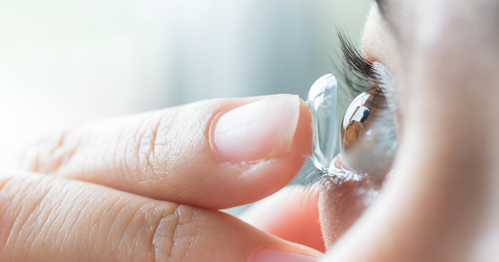
This week’s blog on contact lens wear and care has been contributed by Dr Osama Giledi, Consultant Ophthalmologist, Specialist in Cataract, Cornea and Refractive Vision Correction Surgery.
Here are some useful tips on contact lens wear and care to help you get the best out of your contact lenses.
Things to do:
1. Wear only the lenses specified by your contact lens practitioner
2. Have regular eye check-ups as advised by your ophthalmologist
3. Apply your lenses before putting on and removing make-up
4. Always wash and dry your hands prior to handling your contact lenses
5. Always rub, rinse and store your lenses in the recommended solution before and after each use
6. Discard single-use lenses after each wear
7. Always clean the lens case with solution, wipe with a clean tissue and air dry after each use by placing the case and lids face down on a tissue
8. Carefully handle the lens and check that it is not damaged or inside out before inserting
9. Avoid spraying hairspray or other aerosols into your eye and keep your eyes closed when using any spray
10. Discard lenses and solutions that are past their expiry date
11. Replace your lens case at least monthly
12. Strictly follow the recommended lens wearing schedule and replacement frequency
13. Keep a pair of spectacles with you as backup for when you need to remove your lenses
Always ask yourself each time you are going to wear your contact lenses:
1. Do my eyes feel good with my lenses (no discomfort)?
2. Do my eyes look good (no redness)?
3. Do I see well (no unusual blurring with either eye)?
If the answer to any of these questions is no, then do not wear your contact lenses and consult your contact lens practitioner immediately
Things you should not do:
1. Use tap water, or any other water, on your lenses or lens case
2. Wet your lenses with saliva
3. Wear your lens if it falls on the floor without cleaning it again
4. Wear dirty or damaged lenses
5. Continue to wear your lenses if your eyes don’t feel normal, such as blurred vision or any discomfort
6. Use any eye drops without first consulting your doctor or contact lens practitioner
7. Re-use or top up cleaning solution – discard and replace with fresh solution each time lenses are stored
8. Wear lenses that are left in the case for more than one week without first cleaning and storing them in fresh solution
9. Wear your lenses when showering unless you keep your eyes firmly closed
10. Sleep in your lenses unless advised to by your practitioner and whilst wearing special contact lenses
11. Use your lenses for swimming or water sports, unless wearing protective eye goggles
12. Switch the cleaning solution you use without the advice of your practitioner
13. Share contact lenses with other people
Please remember that if you have any questions or concerns about your contact lenses you should consult your contact lens practitioner or your ophthalmologist for advice

This week’s blog on Floaters and Flashes has been contributed by Dr Ammar Safar, Medical Director, Consultant Ophthalmologist and Vitreoretinal Surgeon.
Some people describe seeing little objects of different shapes and sizes occasionally floating in front of them. We call these objects ‘Floaters’ because they seem to float in front of you wherever you look. In fact, these are small fibres located in the vitreous, the jelly-like substance which fills the eye, in front of the retina. It is very common to see these floaters from time to time and they are more likely to be found in near-sighted individuals.
The floaters are usually seen only when looking at a light background such as a white computer screen or blue sky or sea. Typically, the floaters do not increase with time and if they do, it is usually a very slow progression over many years. The only effective treatment is removing the vitreous, through surgery. However, the procedure is rarely performed as floaters are very benign and rarely cause any significant vision loss. They are more annoying than anything else.
There is no other treatment such as drops or medication that will get rid of them. If there is a sudden increase in the number of floaters (5-10 or more new ones appearing) or if there are flashes of light associated with them, then a visit to your ophthalmologist is necessary to check your retina and ensure there are no holes or tears in it.

Diabetes mellitus is now one of the most common epidemics across continents. While it is a common household term, most people don’t understand what exactly it means to have diabetes. A body with diabetes is unable to properly use the energy from the food ingests, especially carbohydrates, thereby affecting various organs and their functioning.
However, what people know even less about diabetes is that if not treated appropriately, the eyes and visual function can take a severe toll, and diabetes can lead to various vision related diseases.
Diabetic Retinopathy
The initial signs of diabetic retinopathy are small. While modern research is able to detect them, they usually go unnoticed. The retina is the structure at the backside of the eye where the light is processed and the image is transmitted to the brain. Starting as small hemorrhagic spots on the retina, they progress to bigger bleeds, tissue swelling, or retinal contraction. Early control of diabetes can control this.
Macular Edema
The macula is responsible for sharp and detailed vision. The swelling due to diabetes can affect the macula, with decreased visual activity as a result. Further, the photoreceptor cells, that are responsible for converting light to signals, can also be damaged. Modern treatment for Macular Edema include injections to the eye or laser.
Traction Retinal Detachment
Traction retinal detachment occurs due to the contraction of the retina. This leads to dark curtains or blind spots in the field of vision, along with bleeding and formation of holes in the retina. This can be taken care of only through surgery and tenuous post-operative care.
Cataract
Cataract is the clouding of the lens, leading to a decreased quality of vision. Process of cataract is accelerated in diabetics.
As such diabetes represents a threat to human health and vision. It requires constant fight and vigilance as the consequence for its negligence can come at surprisingly high costs. Experts seek solutions for early diagnosis and treatment in fostering collaboration among specialist in various fields including internal medicine, diabetology, endocrinology and others. Early referrals from family medicine and general practitioners are of highest importance. But most of all, patient education and willingness to improve one’s own health should stand out among activities and drives to bring patients for early and regular eye exam.
This blog on the side effects of glaucoma eye drops has been contributed Dr. Salman Waqar, Consultant Ophthalmologist in Cataract and Glaucoma Surgery
Glaucoma is a serious eye condition that can lead to blindness if left untreated. It occurs when there is damage to the optic nerve, which is responsible for transmitting visual information from the eye to the brain. One of the primary causes of optic nerve damage in glaucoma is increased pressure inside the eye, also known as intraocular pressure. Glaucoma eye drops are a common treatment option that can help to lower intraocular pressure and prevent further damage to the optic nerve.
Glaucoma eye drops work by either reducing the amount of fluid that is produced in the eye or by increasing the drainage of fluid out of the eye. There are several different classes of eye drops that are commonly used to treat glaucoma, including prostaglandin analogs, beta blockers, alpha agonists, and carbonic anhydrase inhibitors.
Prostaglandin analogs are often the first line of treatment for glaucoma because they are very effective at lowering intraocular pressure and have few side effects. They work by increasing the drainage of fluid out of the eye. Commonly used prostaglandin analogs include latanoprost, bimatoprost, and travoprost.
Beta blockers work by reducing the amount of fluid that is produced in the eye. They are typically used in combination with other types of eye drops to achieve greater reductions in intraocular pressure. Commonly used beta blockers include timolol, betaxolol, and levobunolol.
Alpha agonists work by reducing the production of fluid in the eye and increasing the drainage of fluid out of the eye. They are often used in combination with other types of eye drops as well. Commonly used alpha agonists include brimonidine and apraclonidine.
Carbonic anhydrase inhibitors work by reducing the amount of fluid that is produced in the eye. They are typically used in combination with other types of eye drops. Commonly used carbonic anhydrase inhibitors include dorzolamide and brinzolamide.
It is important to use glaucoma eye drops as prescribed by your eye doctor. Using the drops as directed can help to reduce intraocular pressure and prevent further damage to the optic nerve. However, it is also important to be aware of the potential side effects of glaucoma eye drops. Some common side effects include stinging or burning of the eyes, redness, blurred vision, and changes in eye color or eyelash growth. If you experience any side effects, be sure to discuss them with your eye doctor.
In conclusion, glaucoma eye drops are an effective treatment option for lowering intraocular pressure and preventing further damage to the optic nerve. There are several different types of eye drops available, each with its own benefits and potential side effects. It is important to work closely with your eye doctor to determine the best course of treatment for your individual needs. With proper use and monitoring, glaucoma eye drops can help to preserve your vision and improve your quality of life.

Too small too soon: Little eyes
One of the many things parents of premature babies say to me is, “I never knew something like this could happen and it has never happened to anyone I know, until now.”
It has been estimated that about 15 million premature babies are born each year around the world. Preterm birth is defined as a baby born before 37 weeks and an extremely preterm baby is born under 28 weeks.
Retinopathy of Prematurity (also called ROP) is an eye disease that affects many premature babies. ROP happens when a baby’s retina doesn’t fully develop in the weeks after birth. The retina is the nerve tissue at the back of the eye. As a consequence of premature birth, abnormal blood vessels are formed, which are fragile and can leak, scarring the retina and pulling it out of position. This causes a retinal detachment, which is the main cause of visual impairment and blindness in ROP.
Who is at risk of developing ROP?
Babies with a birth weight of 1250 grams or less and born before 31 weeks gestation are at the highest risk.
Other factors associated with the presence of ROP in a baby include a lack of weight gain, anemia, when there has been a blood transfusion, breathing difficulties or respiratory distress, and when the baby is generally not in good health.
How do I know if my baby has ROP?
The only way to confirm that a baby is developing ROP is by doing a retinal examination, which is carried out by paediatric ophthalmologists skilled at examining a baby’s eyes. This involves dilating the pupil of the baby with drops. The baby might need weekly or fortnightly retinal examinations until whole of the retina is vascularised (stable and healthy blood vessels).
How is Retinopathy of Prematurity ROP treated?
Some cases of ROP are mild and don’t need any treatment. In severe cases of ROP, the risk of retinal detachment is greater and these babies need treatment either by laser or by injection in the eye to prevent blindness caused by a detached retina.
Early intervention is the key to the treatment of ROP and timely screening is crucial to diagnosis. If a baby has ROP, immediate treatment is critical. The disease can develop very quickly and can destroy a baby’s vision if it’s not treated carefully. So, it is very important for parents to take their baby to all of the planned checkups and eye examinations.