
Joint Commission International Accredited
Committed to providing the highest quality of care
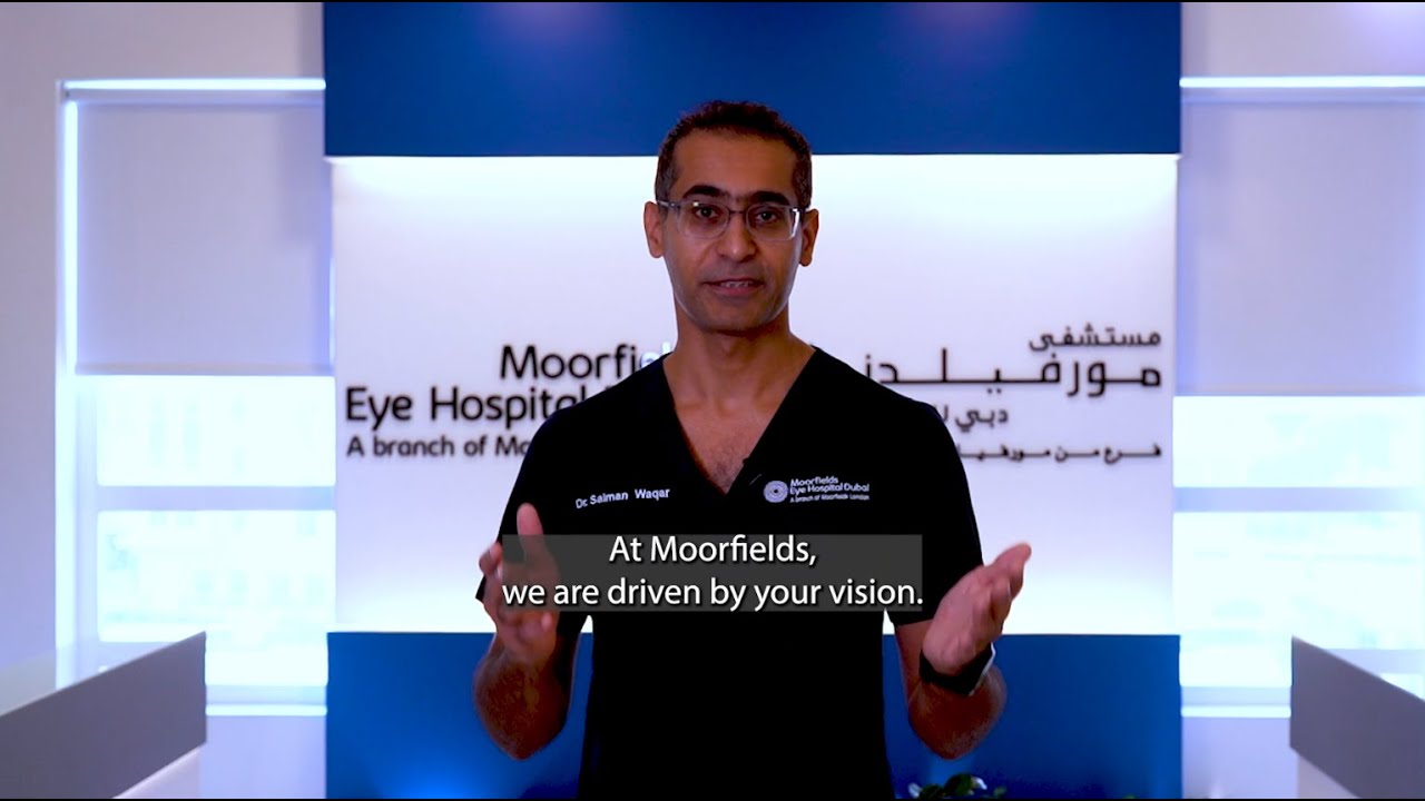
Moorfields Eye Hospitals UAE
200+
15+
350,000+
50+
Driven by your vision
Our Services


Our Specialists
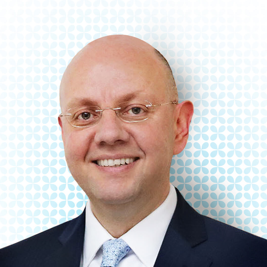
Dr. Ammar Safar
Chief Medical Officer and Consultant Ophthalmologist & Vitreoretinal SurgeonGCAA Approved Specialist Aviation Medical Examiner
Professor of Ophthalmology (Adjunct)
Dr. Ammar Safar is an American Board-Certified Ophthalmologist specialising in vitreoretinal diseases and surgery at Moorfields Eye Hospital Dubai in Dubai Healthcare City.
He is currently an adjunct Professor of Ophthalmology at Mohammad Bin Rashed University and the Chief Medical Officer of Moorfields Eye Hospitals in the United Arab Emirates.
Dr. Safar undertook his medical training in Syria before moving to the United States for post-graduate training. He specialised in Ophthalmology at Georgetown University in Washington, DC, where he also served as a Chief Resident. He then acquired an additional 2-year fellowship training in Vitreoretinal Diseases & Surgery at the University of Texas, Southwestern Medical Center in Dallas, Texas, USA. Dr. Ammar then went on to hold the position of Assistant and later Associate Professor and Director of Vitreoretinal Surgery at the Department of Ophthalmology, Jones Eye Institute, the University of Arkansas for Medical Sciences, in the United States. He then relocated to the UAE, where he was a Consultant Vitreoretinal Surgeon and President of Medical Staff at a private general hospital in Dubai. In addition to his work in private practice, Dr. Ammar has undertaken research and clinical trials in areas such as diabetic retinopathy and age-related eye diseases and published and presented extensively. His research has also resulted in advances in retinal vascular diseases (discovering new management options for diabetic retinopathy and vascular occlusive disorders) and macular degeneration (new approaches for treating exudative macular degeneration).
Among his many honorary awards, Dr. Ammar was the inaugural recipient of The Martha Wood Bentley endowed Chair of Ophthalmology at the University of Arkansas. He is a member of several professional associations, including the American Academy of Ophthalmology, the American Society of Retina Specialists, the European Society of Retina Specialists, the American Society of Cataract and Refractive Surgery, The Association for Research In Vision and Ophthalmology (ARVO), Arab-American Medical Association, Research to Prevent Blindness, and a fellow of the American College of Surgeons.
Dr. Esmaeil Arbabi
Medical Director and Consultant Ophthalmologist in Corneal transplant, Cataract & Refractive SurgeryDr. Esmaeil Mohammad Arbabi is a Consultant Ophthalmologist with excellent surgical experience including all types of corneal transplants (DMEK, DSAEK, PDEK, DALK, PKP), ocular surface reconstruction, complex cataract and laser refractive surgery.
Prior to joining Moorfields Eye Hospital Center, he was Consultant Ophthalmic Surgeon (Cornea, Cataract & Laser Refractive Surgery) and the Lead for Refractive Services at Royal Liverpool University Hospital, Liverpool, United Kingdom.
Dr. Esmaeil holds several prestigious degrees and certifications in the field of cataract and refractive surgery including MSc in Cataract and Refractive Surgery, Certificate in Laser and Refractive Surgery (CertLRS) from Royal College of Ophthalmologist and Fellow of European Board of Ophthalmology Subspecialty Diploma in Cataract and Refractive Surgery (FEBOS-CR).
He is a member of Royal College of Ophthalmologists, European Society of Cataract and Refractive Surgeons, European Board of Ophthalmology, UK Cross-Linking Consortium, EYE -LAW CHAMBERS – Medicolegal Expert and Expert, Association of Personal Injury Lawyers (APIL).
He is instrumental in high quality papers in various peer reviewed journals and numerous scientific and ophthalmology publications. He is also an examiner for the University of Leeds and the University of Liverpool and for the European Board of Ophthalmologist (FEBO exam).
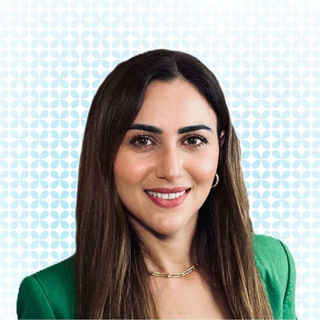
Dr. Alia Issa
Aesthetic Oculoplastic SurgeonDr. Alia Issa is an Aesthetic Oculoplastic Surgeon, specialising in surgical and non-surgical aesthetic eye treatments at Moorfields Eye Hospital Dubai in Dubai Healthcare City.
Dr. Alia is a German board-certified ophthalmologist who completed her medical school and residency training at leading institutions in Germany. She also holds a PhD in Ophthalmology from the University of Dresden, Germany.
She went on to expand her expertise and subspecialise in Aesthetic Oculoplastics by completing a Fellowship training in London at the Ezra Clinic and an Honorary Fellowship in Oculoplastics at the world-renowned Moorfields Eye Hospital in London.
Dr. Alia has over 13 years of regional and international experience in Ophthalmology, including in Germany, the United Kingdom, and the United Arab Emirates.
In her career, she has practised in leading medical institutions such as Moorfields Eye Hospital London, and Schlosspark Hospital Berlin, an academic hospital of the Charite University Hospital Berlin, Gemrany.
She is a member of the German Ophthalmic Society.

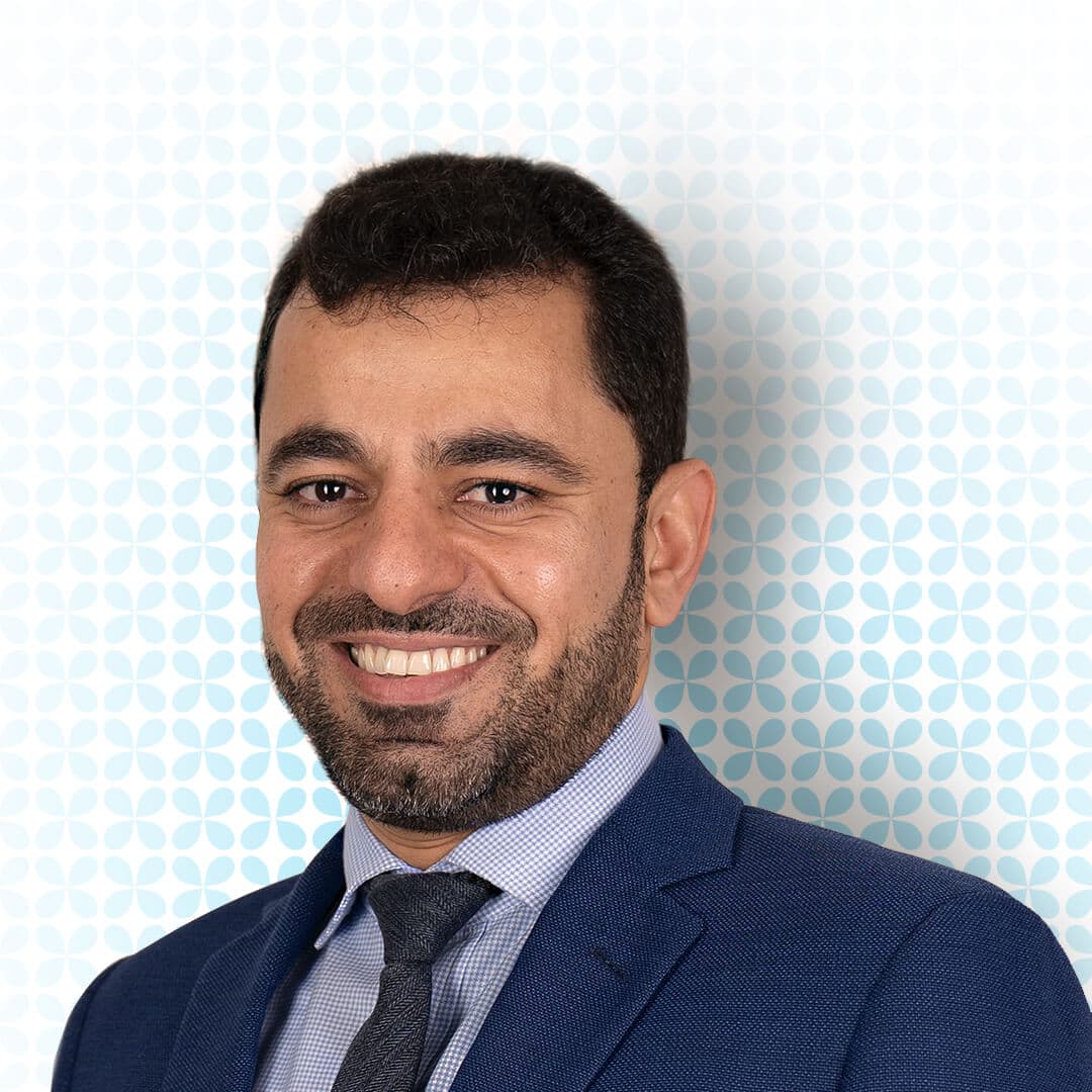
Dr. Alaa A. Mohamed Abou Attawan
Consultant Ophthalmologist in Retina, Uveitis and Diabetic RetinopathyDr. Alaa A. Mohamed Abou Attawan has 15 years of experience in Ophthalmology. He is joining us from Moorfields Eye Centre at Bedford Hospital, United Kingdom, where he was a Consultant Ophthalmologist (Retina). Prior to that, he practiced in Tawam Hospital – Al Ain for 5 years where he set up the foundation and established the Retina service in Al Ain City – UAE.
In his role at Moorfields Eye Hospital Center Abu Dhabi, Dr. Attawan will be working as Consultant Ophthalmologist.
Dr. Attawan graduated from University of Aden, Yemen in 2002 obtaining his Bachelor of Medicine degree. Following his graduation, he moved to the UK to pursue his foundation medical training and further specialty training program in Ophthalmology at the Newcastle upon Tyne Hospitals NHS Foundation Trust and Yorkshire and the Humber School of Ophthalmology working across various hospitals in the UK such as Leeds Teaching Hospitals NHS Trust and Royal Victoria Infirmary from 2008 till 2015. Upon completion of his training, Dr Attawan was awarded the Certificate of Completion of Training (CCT).
He was also a Vitreo Retinal Surgery Fellow at Nottingham University Hospitals, UK from 2015 till 2016.
Dr. Attawan has been a Member since 2008 and a Fellow of the Royal College of Ophthalmologists since 2014. Dr Attawan was a former associate Tutor of the Royal College of Ophthalmologist in London and is actively involved in teaching and research. He has 10 Publications in various prestigious journals and is a well-known speaker at national and regional levels.
He was also one of the Top 10 consultant nominated by SEHA 2022 and he was the Retina service lead and co-founder of Retina service in Al Ain city UAE
Dr. Attawan’s scope of practice covers Cataract surgery, Diabetic eye disease, Retinal Vein occlusion, age-related macular degeneration, emergency eye trauma, and surgery. primary and complex Retinal detachment surgery. Macular hole and Membrane peel surgery.
Dr. Alaa is a Fellow of the Royal College of Ophthalmologists (FRCOphth).
Dr. Fahd Quhill
Consultant Ophthalmologist in Medical Retina and Ocular inflammatory disease (Uveitis)Clinical Professor Of Ophthalmology (Adjunct)
Dr Fahd Quhill is a Consultant Ophthalmologist specialising in Medical Retina and Ocular inflammatory disease at Moorfields Eye Hospital Dubai in Dubai Healthcare City.
Dr Fahd specialises in the assessment and management of uveitis and inflammatory eye diseases (uveitis, infectious diseases, autoimmune diseases of the eye) as well as medical retinal disease, such as diabetes, macular degeneration, inherited retinal disease and retinal vascular disease.
He has broad experience in the diagnosis and management of complex inflammatory eye diseases, including the use of immunosuppression, as well as in treating retinal disease.
Dr. Fahd trained in Ophthalmology in the United Kingdom at Oxford, Birmingham and London, completing three Medical Retinal Fellowships in Birmingham, Wolverhampton and Moorfields Eye Hospital, London.
He has undertaken research and served as principal investigator in numerous macular degeneration, retinal vein occlusion and diabetic macular oedema trials. He has published 27 research papers on retinal and ocular inflammatory disease in leading journal/s such as Ophthalmology, British Journal of Ophthalmology and Eye, among others.
He is a member of several professional bodies, including the Royal College of Ophthalmologists.
Dr. Imad Hakim
Consultant Ophthalmologist in Cataract and Refractive vision correction surgeryDr. Imad Hakim is a Consultant Ophthalmologist, specialised in Cataract and Refractive vision correction surgery at Moorfields Eye Hospital Dubai in Dubai Healthcare City.
He holds a graduate degree from the University of Aleppo in Syria, obtained in 2002. Dr. Hakim also holds a certificate in Ophthalmology from Ludwig Maximillian University of Munich, Germany. He undertook his sub specialty training in Cataract and Refractive surgery at Realeyes Eye Clinic in Munich, Germany.
His areas of expertise are in Femto laser assisted Cataract Surgery (FLACS), Refractive vision correction surgery (LASIK, LASEK, TransPRK, PTK, Implantable Contact Lenses ICLs, Multifocal Intraocular Lenses IOLs and Add-On IOL ), Keratoconus, Phototherapeutic Keratectomy (PTK) Laser Eye Surgery and General Ophthalmology.
He has more than 15 years of regional and international experience in countries including Germany and UAE.
In his career, he has practiced in leading medical institutions such as Realeyes Eye Clinic in Munich, Germany as well as American Hospital in Dubai, U.A.E.
He has published research papers on Cataracts and Retina in leading journals.
He is a member of the European Society of Cataract & Refractive Surgeons (ESCRS), The German Committee for Refractive Surgery (KRC), Commission of the German Ophthalmological Society (DOG) and Professional Association of German Ophthalmologists (BVA).
Dr. Imran Jawaid
Consultant Ophthalmologist in Paediatric Ophthalmology and Strabismus surgeryDr. Imran Jawaid is a Consultant Ophthalmologist, specialised in Paediatric Ophthalmology and Strabismus, at Moorfields Eye Hospital Dubai in Dubai Healthcare City.
He holds a Bachelor of Medicine, Bachelor of Surgery (MBChB) degree in Medicine from the University of Leicester, School of Medicine, graduating with honours in 2010. He is also dual qualified as an Optometrist with a Bachelor of Science (HONS) degree in 2003. Dr. Imran has completed his Fellowship with the Royal College of Ophthalmologists (FRCOphth) and obtained his Certificate of Completion of Training (CCT) in Ophthalmology in 2020.
Dr. Imran has more than 15 years of experience in Ophthalmology in the United Kingdom and has completed a post-CCT fellowship in Paediatric Ophthalmology and Strabismus at Birmingham Children’s Hospital, UK. Most recently he was a Consultant Ophthalmologist and Head of Service for Ophthalmology at Nottingham University Hospitals, NHS Trust, a large tertiary centre for ophthalmic referrals.
He has undertaken research in myopia, visual impairment, diabetic macular oedema and emergency ophthalmology, amongst other areas and has published over 10 research papers in leading journals. In addition to his research, Dr. Imran is a keen educator and has taught at different UK medical schools and contributed to book chapters in Ophthalmology.
He is a member of the Royal College of Ophthalmologists (FRCOphth) and is the recipient of several internationally acclaimed awards and recognitions.
Dr. Luisa M. Sastre
Consultant Ophthalmologist in Medical Retina and Cataract SurgeryGCAA Approved Specialist Aeromedical Examiner
Dr. Luisa Sastre is a Consultant Ophthalmologist in Medical Retina and Cataract Surgery at Moorfields Eye Hospital Dubai in Dubai Healthcare City.
Dr. Luisa holds a Spanish medical degree from the Universidad Autonoma Medical School and completed her ophthalmology residency at Hospital Universitario Gregorio Maranon in Madrid, Spain . She is board certified in Intensive Care medicine and ophthalmology. She also holds a PhD from Universidad Autonoma de Madrid and a Master’s degree in Healthcare Organisation Leadership from ESADE Business School.
She has more than 20 years of combined regional and international experience in countries including Spain and UAE.
She has practised in leading medical institutions such as Hospital Universitario Fundación Jiménez Díaz and Hospital Universitario El Escorial, Madrid, Spain.
She has presented various papers in national and international congresses and journals and organised educational courses on cataract surgery and medical retina for peers and opticians.
She is a member of “Surgical Eye Expeditions International”, a nonprofit humanitarian organisation that hosts clinic sites worldwide. She is also a member of Sociedad Española de Oftalmologia (SEO).
In addition to her clinical work, Dr. Luisa volunteers in various charitable campaigns worldwide to treat patients suffering from cataracts who otherwise have difficulty accessing medical care.
Dr. Miguel Morcillo
Consultant Ophthalmologist in Cornea, Cataract and Refractive vision correction surgeryDr. Miguel Morcillo is a Consultant Ophthalmologist specialising in Cornea, Cataract and Refractive (Vision Correction – LASIK, LASEK, TransPRK) Surgery at Moorfields Eye Hospital Dubai in Dubai Healthcare City.
Dr. Miguel holds a Spanish Degree in Medicine from the Universitat de Valencia. He completed his Residency in Ophthalmology under the MIR (Medico Interno Residente) system of the Spanish Ministry of Health. He has more than 25 years of experience, most of which was attained in Spain. He also worked in Italy and finally moved to the UAE in 2017 and has been practising in the UAE since that time. Dr. Miguel started his career working with Clinica Baviera, the largest private ophthalmology group in Europe, in Madrid and Valencia. He helped develop many of the refractive treatments offered today, such as LASIK, LASEK, ICLs and multifocal IOLs, in addition to collaborations with other hospitals to provide high-quality cataract and refractive surgery to patients. Dr. Miguel trained in cornea surgery at NIIOS Eye Institute (Rotterdam) and, since then became an experienced educator for other surgeons in the fields of the anterior segment and refractive surgery.
He has been a member of “Surgical Eye Expeditions International” since 1999, an American non-government organisation that arranges eye camps worldwide. Dr. Miguel has assisted several eye charity camps in numerous countries of Africa, in addition to Peru, Panama and India, providing free eye care and performing surgeries to those who could not afford it in less developed areas.
His research efforts include surgical topics and outcomes such as Ultrathin LASIK, Keratoconus screening, ICLs, and multifocal IOLs, among others. He has presented various papers in national and international congresses and journals and organised educational courses on refractive surgery for peers and opticians.
He is a member of Sociedad Española de Oftalmologia (SEO), the Panamerican Association of Ophthalmology (PAAO) and the European Vitreoretinal Society (EVRS).
He is also the recipient of the Dr. P C Mahendra Memorial ACOIN International Award in India in recognition of his charitable work with people in need.
Dr. Nadim Habash
Consultant Ophthalmologist in Cornea and Ocular Surface DiseasesDr. Nadim Habash is an American Board-Certified Consultant Ophthalmologist specialising in Cornea, Ocular Surface Diseases, and General Ophthalmology at Moorfields Eye Hospital Dubai in Dubai Healthcare City.
Dr. Nadim undertook his speciality training at George Washington University in Washington D.C., U.S.A., where he also served as Chief Resident. He then acquired an additional Cornea and External Disease Fellowship at the University of Florida, Gainesville, U.S.A.
Dr. Nadim is a Diplomate of the American Board of Ophthalmology and a Fellow of the Royal College of Surgeons of Canada FRCS(C).
Over the past 40 years, Dr. Nadim has had extensive experience in Corneal transplantation and Anterior Segment Diseases in several countries, including the United States of America, the Kingdom of Saudi Arabia and the United Arab Emirates.
Dr. Nadim has worked and held leadership and academic positions in prestigious institutions, including the University of Ohio at Toledo, Ohio, U.S.A., the King Faisal Specialist Hospital and the King Khaled Eye Specialist Hospital in Saudi Arabia, and at the American Hospital Dubai, where he held key positions including Chief of Medical Staff, Assistant Chief Medical Officer and Director of Ophthalmology.
Dr. Nadim published several papers about Corneal transplantation, chaired many scientific conferences and symposia, and participated actively in teaching medical students and Ophthalmology residents.
Throughout his career, Dr. Nadim held memberships in several scientific organisations, including a fellowship of the American College of Surgeons, the American Academy of Ophthalmology membership, and many regional and local societies
Dr. Osama Giledi
Consultant Ophthalmologist in Cataract, Cornea and Refractive Vision Correction SurgeryGCAA Approved Specialist Aeromedical Examiner
Associate Professor Of Ophthalmology (Adjunct)
Dr. Osama Giledi is a Consultant Ophthalmologist specialising in Cornea, Cataract and Refractive Surgery at Moorfields Eye Hospital Dubai in Dubai Healthcare City.
Dr. Osama is skilled in managing ocular surface problems, including severe dry eye and Stem cell deficiency. He performs small incision phacoemulsification for his cataract surgery and is experienced in using toric and multifocal premium intraocular lenses. Dr. Osama’s expertise in managing complex corneal conditions includes all types of modern corneal graft procedures, such as DALK and DSAEK. He has performed over 23,000 refractive surgeries, including Lasik, LASEK, Intralase LASIK and Trans PRK, and phakic IOLs. He delivers the latest treatment for keratoconus, including Intracorneal ring segments, corneal cross-linking and complex laser treatment.
Dr. Osama graduated from Libya and completed his ophthalmic training in the UK, attaining a Fellowship in Ophthalmology from The Royal College of Edinburgh in 1996. He completed two years of higher subspecialty training fellowship on the anterior segment, Cornea and refractive surgery in 2003 at the prestigious Corneoplastic Unit and Eye Bank at Queen Victoria Hospital, East Grinstead. He worked as a Consultant Ophthalmologist at the Centre for Sight London and the Corneoplastic Unit and Eye Bank at Queen Victoria Hospital. Dr Osama has 22 years of experience in the UK, providing services for anterior segment, Cornea, Refractive and cataract surgery.
In addition to his clinical commitments, Dr. Osama has extensive experience in teaching and training; he is a noted presenter at national and international meetings and has extensive research published in peer-reviewed scientific journals. He is a member of the Royal College Surgeons of Edinburgh, the United Kingdom & Ireland Society of Cataract and Refractive Surgeons, and the European Society of Cataract and Refractive Surgeons.
Dr. Rafic Antonios
Specialist Oculoplastic SurgeonDr. Rafic Antonios is a fellowship-trained Oculoplastic surgeon with expertise in reconstructive and cosmetic eye procedures involving the periocular area and pathologies involving the eyelid (Blepharoplasty), orbit, and lacrimal system at Moorfields Eye Hospital Dubai in Dubai Healthcare City.
He holds a Bachelor of Science (2008) with distinction and a Medical Degree (2012) from the American University of Beirut. He finished his ophthalmology residency at the American University of Beirut Medical Center in (2017) with induction to the Alpha Omega Alpha (AOA) – Honor Medical Society before pursuing a fellowship in Oculoplastics and Reconstructive Surgery and an additional fellowship in craniomaxillofacial trauma at the University of Montreal, Canada (2019).
He has over five years of regional and international experience in ophthalmic plastic and reconstructive surgery in Canada and Lebanon.
Dr. Rafic has practised in leading medical institutions such as Saint Justine Children’s Hospital, Maisonneuve-Rosemont Hospital, Sacre-Coeur Hospital in Canada, and the Beirut Eye & ENT Specialist Hospital in Lebanon.
He has 28 publications in his field in peer-reviewed journals and several posters and presentations at national and international conferences.
He is a member of the Alpha Omega Alpha (AOA) Medical Honor Society and the Lebanese Society of Ophthalmology (LOS).
Dr. Ramzi Ghanem
Consultant Ophthalmologist in Medical Retina, Vitreoretinal and Cataract SurgeryDr. Ramzi Ghanem is a Consultant Ophthalmologist with extensive experience in cataract surgery as well as medical and surgical retina including the full spectrum of vitreoretinal surgery, intravitreal drug injection and laser treatment.
Dr. Ramzi is a graduate of the University of Leipzig, Germany and completed his 2 doctoral thesis on anti-VEGF isomers in AMD (Age-related Macular Degeneration) at the University of Leipzig and predictors for short term visual outcome after anti-VEGF therapy of CSME due to CRVO at the University of Bern, Switzerland. He has more than 20 years of experience in Germany, Switzerland and UAE.
He has researched extensively, conducted clinical trials, published and lectured on his areas of expertise. He has been a HAAD consultant and examiner for licensure in ophthalmology.
Dr. Salma Yassine
Specialist Ophthalmologist in Paediatric and Neuro-ophthalmologyDr. Salma Yassine is a specialist ophthalmologist in paediatric and neuro-ophthalmology at Moorfields Eye Hospital Dubai in Healthcare City Dubai.
She holds a bachelor’s in science (2012) with high distinction and a medical degree (2016) from the American University of Beirut. She finished Her ophthalmology residency at the American University of Beirut Medical Center in 2020 before pursuing a fellowship in neuro-ophthalmology at the University of Minnesota in 2021, followed by another fellowship in pediatric ophthalmology and strabismus at Children’s National Medical Center in Washington DC.
She has international experience in paediatrics and neuro-ophthalmology in countries including the USA and Lebanon.
She has practised in leading medical institutions such as the University of Minnesota and the Children’s National Medical Center in the US.
She has researched intracranial hypertension and nanophthalmos and published research papers on nystagmus and nanophthalmos in leading journals.
She is a member of the American Association for Pediatric Ophthalmology and Strabismus Association (AAPOS) The North American Neuro-Ophthalmology Society (NANOS), and the Lebanese Ophthalmological Society (LOS)
Dr. Salman Waqar
Consultant Ophthalmologist in adult and paediatric glaucoma and cataract surgeryAssociate Professor Of Ophthalmology (Adjunct)
Dr. Salman Waqar is an experienced consultant ophthalmologist specialising in the treatment of adult and paediatric glaucoma and cataracts at Moorfields Eye Hospital Dubai in Dubai Healthcare City.
His areas of expertise include medical, laser and surgical treatments for glaucoma (including minimally invasive implants and aqueous shunt devices). He also offers cataract surgery using the latest small incision technology, and with a range of intraocular lenses (Toric, Multifocal and Extended Depth of Field), tailored to the patients individual needs.
Dr. Waqar graduated from Pakistan and has over 15 years’ experience of work in the United Kingdom. He holds a Certificate of Completion of Training from the UK, and is a Fellow of the Royal College of Ophthalmologists. He has worked in various prestigious eye units including the West of England Eye Unit (Exeter), where he completed his sub-specialty training in glaucoma. In his career, he has been appointed as a consultant across a wide range of leading medical institutions such as the Royal Eye Infirmary (Plymouth), Nuffield Health, Medical Eye Clinic and Newmedica. In addition to his clinical work, he has also performed with distinction as director of clinical governance and for the glaucoma service.
Dr. Waqar’s research interests include optimizing patient outcomes with minimally invasive surgery, using virtual reality simulation to investigate surgeon performance and the development of innovative surgical instruments to improve patient safety. He has published extensively in peer-reviewed journals, such as the British Journal of Ophthalmology and the Journal of Cataract and refractive Surgery, and presents regularly at national and international meetings.
He holds memberships with the United Kingdom and Eire Glaucoma Society, the European Society of Cataract and Refractive Surgeons and the American Academy of Ophthalmology.
In his capacity as an Honorary Fellow with Plymouth University, he has been involved in training the doctors and surgeons of our future. In 2019, his work was recognized with a nomination for a regional Trainer of the Year Award by the Peninsula School of Ophthalmology. In addition, he has been the recipient of numerous research grants, and has co-authored two medical textbooks. Other accolades include invitations as a surgical skills instructor at minimally invasive glaucoma surgery courses, and to the microsurgical skills course with the Royal College of Ophthalmologists.
In his spare time he is an avid reader, aspiring author and motivated triathlete.
Dr. Suhair Twaij
Consultant Ophthalmologist in Adult & Paediatric Strabismus Surgery, Paediatric Ophthalmology, Adult Cataract SurgeryDr. Suhair Twaij is a Consultant Ophthalmologist with a subspecialist interest in adult and paediatric Strabismus surgery, Paediatric Ophthalmology, adult Cataract Surgery and General Ophthalmology at Moorfields Eye Hospital Dubai.
Dr. Twaij recently relocated to the UAE from the UK, where she has worked as a consultant ophthalmologist at the Royal Victoria Hospital in Belfast since 2013. She was also the clinical lead for the adult motility service in Northern Ireland from 2014.
She holds a graduate degree in medicine from the Baghdad University Medical School, obtained in 1994.
Her areas of expertise include complex adult and paediatric squint surgery using the adjustable technique when needed, double vision correction, adult cataract surgery with an implant, Botox treatment for squints, double vision, blepharospasm and aesthetics.
She has over 20 years of experience in general ophthalmology, including cataract surgery, anterior and posterior segments such as glaucoma and diabetic retinopathy.
Dr. Suhair trained in the UK from 2004 in England and Northern Ireland and was awarded the certificate of completion of training (CCT) in ophthalmology in the UK in 2012. She was awarded the Royal College of Ophthalmologists Fellowship in the same year.
Throughout her career, she has practised in leading medical institutions such as The Royal Victoria Hospital in Belfast, Northern Ireland, Altnagelvin Area Hospital, Northern Ireland, The Bristol Eye Hospital, England, and King Hussein Medical City Amman, Jordan.
Dr. Suhair is an avid researcher and has Medline-published papers in various ophthalmology topics such as medical retina, paediatric ophthalmology and oculoplastics.
She is a member of the Royal College of Ophthalmologists, the British & Irish Paediatric Ophthalmology and Strabismus Association (BIPOSA) and the European Association of Strabismus (ESA).
Dr. Syed M. Asad Ali
Consultant Ophthalmologist in Paediatric Ophthalmology and Strabismus and Cataract SurgerySyed Ali is a Consultant Paediatric Ophthalmologist with extensive experience in the management of all paediatric eye conditions including congenital eye diseases, paediatric cataracts, paediatric glaucoma, trauma and all types of squints in children and adults.
Syed works with advanced technology and techniques. He has over 10 years of extensive experience in screening and treating retinopathy of prematurity with retinal laser and intravitreal anti VEGF injection. He has wealth of experience in phacoemulsification procedures.
Syed Ali trained and has practiced in the UK and Pakistan and has over 18 years of experience in ophthalmology. After completing his basic specialist training, he undertook a fellowship in paediatric ophthalmology and ocular motility at the world renowned Wolverhampton Eye Infirmary, UK. His training in Pakistan and the UK is recognised by the General Medical Council (GMC) and was placed on specialist register.
In addition to his clinical experience, Syed has undertaken published original research and teaching in his specialist area, including as an assistant professor of ophthalmology at the Pakistan Institute of Ophthalmology.
He is a member of several professional bodies including The Royal College of Ophthalmologists, American Academy of Ophthalmology, The British Medical Association, Medical Defense Union and Royal College of Surgeons Edinburgh.
Dr. Irfan Khan
Consultant Ophthalmic surgeon in Paediatric Ophthalmology and adult StrabismusDr. Muhammad Irfan Khan is a highly experienced consultant Ophthalmic surgeon specialising in Paediatric Ophthalmology at Moorfields Eye Hospitals in Abu Dhabi and Dubai.
He earned three postgraduate degrees from the Royal College of Ophthalmologists London: DRCOphth, MRCOphth, and FRCOphth in 2013, along with a Paediatric Ophthalmology fellowship from the renowned Hospital for Sick Children, SickKids, Toronto, Canada.
With over 20 years of clinical and surgical experience, Dr. Irfan has practised in several countries, including Ireland, the United Kingdom, Canada, and the UAE. His career includes roles at institutions such as the Royal Victoria Eye and Ear Hospital in Dublin, Manchester Royal Eye Hospital, SickKids in Toronto, and Sheikh Khalifa Medical City in Abu Dhabi.
His subspecialty training includes managing complex paediatric eye conditions such as cataracts, glaucoma, and retinopathy of prematurity. He is also experienced in paediatric anterior segment reconstruction and common paediatric oculoplastics.
For adult patients, Dr. Irfan is particularly adept in performing adjustable suture squint surgeries, a technique that allows for precise postoperative alignment adjustments. This approach significantly improves outcomes for adults undergoing strabismus surgery, enhancing both visual function and cosmetic results.
He has undertaken research in the field of Ophthalmology and has published ten research papers and one book chapter in Childhood neurology.
He is a member of the Royal College of Ophthalmologists London and an International member of the American Association of Pediatric Ophthalmology and Strabismus. In 2015, he received the JD Moran Award from the Hospital of Sick Children in Toronto, Canada.
Dr. Ithar Alkheder
Specialist general OphthalmologistDr. Ithar Alkheder is a distinguished Specialist Ophthalmologist at Moorfields Eye Hospital Dubai, located in Dubai Healthcare City. His areas of expertise are general ophthalmology and comprehensive eye examinations.
He earned a Master’s degree in Ophthalmology and holds board certification from the Syrian Board Authority since 2011. His qualifications are further complemented by a Diploma in Pediatric Optics from Isabel University, Spain.
Dr. Alkheder brings over 13 years of regional and international experience, having practiced in several medical institutions both the UAE and Syria.
His research pursuits include corneal ulcer diagnosis and treatment at Aleppo Eye Surgery Hospital. He has published various research papgers in leading medical journals and presented in international ophthalmology conferences, focusing on penetrating glaucoma surgery, among other topics.
Dr. Alkheder is a member of the American Academy of Ophthalmology (AAO), the European Society of Retina (Euretina), and the International Society of Refractive Surgery (ISRS), reflecting his deep commitment to his field and continuous professional development.
Dr. Alaa Bou Ghannam
Visiting Specialist Ophthalmologist in Neuro-Ophthalmology, Paediatric Ophthalmology, Paediatric Glaucoma & Adult StrabismusDr. Alaa Bou Ghannam is a visiting specialist in neuro-ophthalmology, paediatric ophthalmology, and adult strabismus at Moorfields Eye Hospital Dubai in Dubai Healthcare City.
He holds a bachelor in science (2007) with high distinction and a medical degree (2011) from the American University of Beirut. He finished his ophthalmology residency at the American University of Beirut Medical Center in 2015 before pursuing a fellowship in pediatric ophthalmology and adult strabismus from Children’s National Health System and George Washington Hospital in Washington DC in 2016. He also finished another fellowship in neuro-ophthalmology from The University of Colorado in 2017.
He has over five years of regional/international experience in paediatric ophthalmology, adult strabismus, and neuro-ophthalmology in countries including Lebanon and The United States of America.
In his career, he has practiced in leading medical institutions such as The American University of Beirut Medical Center.
He has undertaken research in clinical ophthalmology and neurology and has published more than a dozen research papers and book chapters on strabismus, optic nerve diseases, paediatric glaucoma, paediatric ophthalmology, ocular electrophysiology, and nystagmus, in leading medical journals.
He is a member of the American Academy of Ophthalmology (AAO), Lebanese Ophthalmological Society (LOS), North American Neuro-ophthalmology Society (NANOS), and The American Association for Pediatric Ophthalmology and Strabismus (AAPOS).

Dr. Avinash Gurbaxani
Visiting Consultant Ophthalmologist in Uveitis and Medical Retinal Diseases and Cataract SurgeryDr. Avinash Gurbaxani is a visiting Consultant Ophthalmologist, specialised in Uveitis, Medical Retina & Cataract surgery at Moorfields Eye Hospital Dubai in Dubai Healthcare City.
Dr. Avinash specialises in the assessment and management of uveitis and inflammatory eye disease (uveitis, infectious diseases, autoimmune diseases of the eye) as well as medical retinal disease, such as diabetes, macular degeneration and retinal vascular disease. He has broad experience in the diagnosis and management of complex inflammatory eye diseases, including the use of immunosuppression, as well as in treating retinal disease. Dr Gurbaxani is also skilled in managing complex cataract surgery associated with these diseases.
Dr. Avinash trained in ophthalmology in Oxford and London, working at The Oxford Eye Hospital in Oxford, Kings College Hospital, St. Thomas’ Hospital and Moorfields Eye Hospital in London. He has worked at the prestigious Medical Eye Unit in London as well as completing a Uveitis Fellowship and Medical Retina Fellowship at Moorfields London and the Sydney Eye Hospital in Australia, before returning to Moorfields London as a locum consultant.
Dr. Avinash is experienced in initiating research projects and running clinical trials, and has regularly published and presented papers at national and international conferences. He is a member of several professional bodies including the Royal College of Opthalmologists (RCOphth), Royal College of Surgeons of Edinburgh (RCS) (Ed), American Academy of Ophthalmology (AAO).
Dr. Christiane Al Haddad
Visiting Consultant Ophthalmologist in Paediatric Ophthalmology and Adult StrabismusDr Christiane Al- Haddad is a visiting associate professor of Ophthalmology, Specialised in Paediatric Ophthalmology and adult Strabismus at Moorfields Eye Hospital Dubai in Dubai Healthcare City.
She holds an MD degree from the American University of Beirut in 2000, an Ophthalmology degree from the American University of Beirut in 2004, a fellowship in paediatric ophthalmology from Children’s National Medical Center, Washington DC in 2005, and a fellowship in paediatric ophthalmology and adult strabismus from Duke Eye Center, Durham, North Carolina, USA in 2006.
Dr Al-Haddad has more than 15 years of regional/international experience in all aspects of Paediatric Ophthalmology and Strabismology in leading medical institutions: the American University of Beirut Medical Center (Lebanon), Children’s National Medical Center (USA) and Duke Eye Center (USA).
She has undertaken research in the field of Paediatric Ophthalmology & strabismus and has published more than 50 research papers on amblyopia, strabismus, genetic eye diseases, retinoblastoma, congenital glaucoma and eye tracking technology in leading journals including British Journal of Ophthalmology, American Journal of Ophthalmology, Journal of the American Association for Paediatric Ophthalmology and Strabismus, Journal of Paediatric Ophthalmology and Strabismus, and Canadian Journal of Ophthalmology among others.
Dr Al-Haddad is a member of the American Association for Paediatric Ophthalmology and Strabismus, the International Strabismological Association and the Costenbader Society. She is the director of the Paediatric Ophthalmology division in the Ophthalmology department at the American University of Beirut and established the retinoblastoma program with the oncology team at the Children’s Cancer Center in collaboration with St Jude Children’s Research Hospital, Memphis USA.
Dr. Khalid Alkhayat
Visiting Consultant Ophthalmologist in Oculoplastic SurgeryDr. Khalid Alkhayat is a Visiting Consultant Ophthalmologist, specialised in Oculoplastics at Moorfields Eye Hospital Dubai in Dubai Healthcare City.
He is a German Board Certified Ophthalmologist (Fachartz) and Fellow of the European Board of Ophthalmology (FEBO) since 2017.
Dr. Alkhayat has over 12 years of combined regional and international experience in Oculoplasty in countries including the UAE, Ireland and Germany.
During his career, he has practiced in leading medical institutions such as the Royal College of Surgeons in Ireland, Bruder Krankenhausin Trier, Germany, Gulf Medical University in Ajman, UAE, Dubai Hospital, UAE, Rashid Hospital, Dubai, UAE and Al Mafraq Hospital in Abu Dhabi, UAE.
He is also a member of Emirates Society of Ophthalmology (ESO).
Dr. Qasiem Nasser
Visiting Consultant Ophthalmologist in Oculoplastic SurgeryDr. Qasiem Nasser has dual Fellowship training in Oculoplastic Surgery and Cornea, Cataract and Anterior Segment Surgery. His area of expertise is in Ophthalmic Plastic and Reconstructive Surgery, including surgery on the eyelids, the orbit, and the tear drainage system for adults and children. In addition, he is also a specialist in Laser Assisted Cataract and Refractive Surgery, including LASIK, LASEK, Refractive Lens Exchange (RLE), Implantable Contact Lens (ICL) and Corneal Cross-Linking (CXL) for Keratoconus.
Dr. Nasser grew up in Amman, Jordan. He attended medical school at The Royal College of Surgeons in Ireland, after which he completed his Residency in Ophthalmology at The Royal Victoria Eye and Ear Hospital in Dublin. He then moved to the United States, where he completed a 2-year Ophthalmic Plastic and Orbital Reconstructive Surgery Fellowship at The University of Texas MD Anderson Cancer Center in Houston and Texas Oculoplastic Consultants in Austin. Dr. Nasser then persuaded another Fellowship in Cornea, Cataract and Anterior Segment Surgery at Moorfields Eye Hospital in London.
He is a member of the Royal College of Ophthalmologists in London and a Fellow of the Royal College of Surgeons in Edinburgh.
Dr. Nasser has been an active researcher and has published many manuscripts in peer-reviewed journals. In addition, he has given lectures at national and international meetings, including The American Society of Ophthalmic Plastic and Reconstructive Surgery, the American Academy of Ophthalmology and the World Ophthalmology Congress.
Dr. Rola Ba-Abbad
Visiting Consultant Ophthalmologist Specialised in Genetic Eye Diseases and Medical RetinRola Ba-Abbad is a Consultant Ophthalmologist in genetic eye disease, including inherited retinal disorders for adults and children, and medical retina at Moorfields Eye Hospital Dubai in Dubai Healthcare City.
She holds a PhD from University College London, 2018, and is a Fellow of the Royal College of Physicians and Surgeons of Glasgow since 2011. She earned her bachelor’s degree (MBBS) in Medicine and Surgery from King Saud University and successfully completed her specialist training at King Khaled Eye Specialist Hospital and affiliated hospitals in Riyadh in 2006.
She has more than 10 years of regional and international experience in medical retina, electrophysiology and inherited retinal disorders in countries including Saudi Arabia, the United Kingdom and Canada.
In her career, she has practised in leading medical institutions such as Moorfields Eye Hospital in London, Hospital for Sick Children in Toronto, and King Khaled Eye Specialist Hospital.
She has undertaken research in inherited retinal disorders, visual neuroscience-clinical psychophysics, and electrophysiology of vision and has published several research papers on retinal and corneal disorders in leading journals such as Ophthalmology and Investigative Ophthalmology and Visual Sciences, among others.
She is a member of The Association for Research in Vision and Ophthalmology and is the recipient of the Foundation Fighting Blindness: Diana Davis Spencer Clinical Research Fellowship Award in 2018.
Dr. Razek Coussa
Visiting Consultant Ophthalmologist in Adult and Paediatric vitreoretinal diseases & surgery, inherited retinal diseases and retinal gene therapyDr. Razek Coussa is a visiting consultant Ophthalmologist specialising in adult and paediatric vitreoretinal diseases and surgery, inherited retinal diseases and retinal gene therapy at Moorfields Eye Hospital Dubai in Dubai Healthcare City.
Dr. Razek academic journey started at McGill University (Montreal, Canada), where he graduated with a Bachelor in Chemical Engineering (Dean’s Honours list) in 2007, followed by a Masters in Biomedical Engineering in 2008. After obtaining a Masters in Philosophy in Bioscience Enterprise from the University of Cambridge (United Kingdom 2009), he returned to McGill University, where He completed his training in Medicine (2009-2013), followed by a post-graduate residency in Ophthalmology (2013-2018). After which he successfully obtained the Canadian Royal College certification in Ophthalmology and the American Board of Ophthalmology certification.
Dr. Razek then completed four years of fellowship training, developing comprehensive clinical, surgical, and research expertise in multiple retina-related sub-specialities. His training included a one-year fellowship in Paediatric Ophthalmology and Inherited Retinal Diseases at the prestigious Cole Eye Institute of the Cleveland Clinic (2018-2019), advanced training in Medical Retina and Molecular Ophthalmology at the world-renowned Department of Ophthalmology of the University of Iowa (2019-2020), and extensive training in Vitreoretinal Diseases and Surgery at Iowa (2020-2022).
Dr. Razek brings over 10 years of regional/international experience in medical and surgical vitreoretinal diseases, and inherited retinal diseases, having worked in countries including the United States and Canada. This global perspective enriches his approach to patient care and research.
In his career, he has practised in leading medical institutions such as the University of Iowa (Iowa City, Iowa, USA), Cole Eye Institute of the Cleveland Clinic (Cleveland, Ohio, USA), The Dean McGee Eye Institute (Oklahoma City, Oklahoma, USA) and McGill University Health Center (Montreal, Quebec, Canada).
Dr. Razek has undertaken research in surgical and inherited retinal diseases and inherited retinal diseases and has published over 50 peer-reviewed articles and book chapters on diabetic retinopathy, vitreomacular traction, Choroideremia and Stargardt disease, among others.
He is a member of the American Academy of Ophthalmology (AAO), the American Society of Retina Specialists (ASRS), the International Society for Genetic Eye Diseases and Retinoblastoma (ISGEDR), and a fellow of the Royal College of Physicians and Surgeons of Canada (RCPSC).
Dr. Wajiha Kheir
Visiting Consultant Ophthalmologist in Medical Retina and Ocular OncologyDr. Wajiha Kheir is a visiting Assistant Professor of Ophthalmology and Consultant Ophthalmologist, specialised in Medical Retina and Ocular Oncology at Moorfields Eye Hospital Dubai in Dubai Healthcare City.
Dr. Wajiha completed her undergraduate studies and medical education at the American University of Beirut, Lebanon. She was awarded the BSc degree in Biology (2009) with high distinction and the Penrose Award, and the MD degree (2013) with induction to the Alpha Omega Alpha (AOA) – Honor Medical Society. She then completed a year of Internal medicine Internship followed by three years of Ophthalmology residency at the American University of Beirut Medical Center (2017). Her subsequent sub-specialty training includes one year of Medical Retina fellowship and one year of Ocular Oncology with Clinical Pathology Concentration fellowship, both at the Duke Eye Center, Duke University School of Medicine.
She has more than five years of regional and international experience in Medical Retinal and Ocular Oncology in the United States and Lebanon. In her career, she has practiced in leading medical institutions such as Duke University Health System and the American University of Beirut Medical Center.
Dr. Wajiha has eighteen peer-reviewed journal articles and 10 book chapters published. To date, her research work has been presented in over twenty posters and/or paper presentations at regional and international conferences. Her research interests relate to age-related macular degeneration, ophthalmic oncology, radiation retinopathy as well as retinal degenerations and electrophysiology of vision.
She is a member of the AOA Medical Honor Society, the American Academy of Ophthalmology, the American Society of Retinal Specialists and the International Society of Ocular Oncology.
Prof. Ziad Bashshur
Visiting Consultant Ophthalmologist in Medical & Surgical RetinaDr. Ziad Bashshur is a visiting Ophthalmologist specialising in Medical and Surgical Retina at Moorfields Eye Hospital Dubai in Dubai Healthcare City.
He holds a Doctor of Medicine Diploma from the American University of Beirut (AUB) in 1994. He has also completed an internship in internal medicine at Georgetown/VA medical center (1994-1995) followed by ophthalmology residency training at AUB (1995-1998) and a vitreoretinal fellowship at the University of Virginia, USA.
He has more than 22 years of regional/international experience in vitreoretinal diseases and surgery in countries including United States and Lebanon.
In his career, he has practiced in leading medical institutions such as American University of Beirut in Lebanon and University of Virginia in the US.
He is currently a tenured professor and vice-chairman of Ophthalmology at the American University of Beirut.
Dr. Bashshur has undertaken research in retinal diseases and has published over 50 research papers on macular degeneration, diabetic retinopathy, vascular diseases of the retina and retinal imaging in leading journals. In addition, he sits on several advisory boards within the medical industry.
He regularly speaks at professional meetings worldwide and serves on the review board of several renowned peer-reviewed ophthalmology journals.
He is a member of the American Society of Retina Specialists (ASRS), Retina Society and Alpha Omega Alpha Honor Society
Dr. Ziad Khoueir
Visiting Consultant Ophthalmologist in Adult Glaucoma and Cataract surgeryDr. Ziad Khoueir is a visiting Consultant ophthalmologist specialising in adult glaucoma and cataract surgery at Moorfields Eye Hospital Dubai in Dubai Healthcare City.
He holds a medical degree from the Saint-Joseph University in Beirut, Lebanon (2008) and completed his ophthalmology residency at the Hotel-Dieu de France Hospital (2013). He then completed postdoctoral clinical and research fellowships at the Glaucoma Institute, UPMC-Sorbonne University in Paris, France (2014) and at the Massachusetts Eye and Ear Infirmary, Harvard Medical School in Boston, USA (2016). He is also a laureate of the Glaucoma Subspecialty Diploma of the European Board of Ophthalmology.
He has more than seven years of both regional and international experience in glaucoma, one during which he has practised clinical and research work in leading medical institutions such as the Glaucoma Institute at the Paris Saint-Joseph Hospital (France), the Massachusetts Eye and Ear Infirmary (USA), the Beirut Eye & ENT Specialist Hospital (Lebanon) and the Muscat Eye Laser Center (Oman).
Dr. Ziad currently holds the position of associate director of the Glaucoma division at the Beirut Eye and ENT Specialist Hospital. He is a clinical instructor and lecturer at the Saint-Joseph University Medical School in Beirut, Lebanon.
His research focuses on innovative techniques in managing and diagnosing glaucoma, such as minimally invasive glaucoma surgery (MIGS), glaucoma lasers and the use of Optical Coherence Tomography imaging in glaucoma. He has published more than 35 research papers and chapters on those topics in leading ophthalmology journals. He has been invited to lecture and present his work in regional and international meetings in more than 20 countries over the last few years. He is currently a research collaborator at the Mayo Clinic Ophthalmology Department in Jacksonville, Florida, USA.
He is the co-chair of the associate advisory board of the World Glaucoma Association and a member of the associate advisory board of the International Society of Glaucoma Surgery.
Dr. Aseel Ghanem
Consultant Ophthalmologist in Medical Retina and Refractive surgeryDr. Aseel Ghanem is a Consultant Ophthalmologist at Moorfields Eye Hospital Abu Dhabi and Imperial College London Diabetes Centre (ICLDC) with more than 8 years of experience. She is experienced in refractive surgeries, medical retina, lacrimal surgeries, pterygium excision with graft, squint surgery, eyelid laceration repair & more.
She completed her medical degree from Mutah University, Jordan in 2012 and completed her residency from Ibn Al Haytham Hospital, Jordan. She is a fellow of International Council of Ophthalmology.
Dr . Usman Mahmood
Consultant, OphthalmologistDr. Usman Mahmood is a Consultant Ophthalmologist with extensive experience in vitreoretinal surgery, sutureless advanced cataract surgery, advanced Diabetic retinopathy, retinal detachment repairs, macular holes, epiretinal membranes and ocular trauma involving posterior segment.
Dr. Usman is a fellow in FRCS (Ophthalmology) Royal college of Surgeons Edinburgh UK, FRCS (Ophthalmology) Royal College of Surgeons and Physicians of Glasgow UK, MRCOphth Royal College of Ophthalmologists London UK. He obtained Certificate of Specialist training in Ophthalmic Surgery from Royal College of surgeons in Ireland. He is a fellow in Vitreoretinal Surgery from Manchester Royal Eye Hospital, Manchester, UK.
Dr. Usman is also a member European Society of Vitreoretinal Surgeons and a Life member of Ophthalmic Society of Pakistan.
Dr. Omar Rafiq
Consultant, Ophthalmologist Specializing in small incision cataract surgery (phacoemulsification) and advanced glaucoma surgeryDr. Omar Rafiq is a Consultant Ophthalmologist specializing in small incision cataract surgery (phacoemulsification) and advanced glaucoma surgery including MIGS ( Minimally Invasive Glaucoma Surgery ). He has performed over 10,000 surgical procedures.
Dr. Omar is a graduate of the University of Leeds Medical School – obtaining his MBChB, from the United Kingdom. He is a Fellow of the Royal College of Ophthalmologists – UK (FRCOphth) and holds a Certificate of Completion of Training (CCT) and is on the General Medical Council Specialist Register for Ophthalmology. Following the CCT, he completed an advanced fellowship in glaucoma and complex cataract at the Western Eye Hospital in London. He also completed his Postgraduate Diploma in Cataract and Refractive Surgery from the University of Ulster with a Distinction grade. He has more than 15 years of experience in the United Kingdom and the United Arab Emirates. Among many other experiences, he was the youngest Director of the NSPCC at a regional level from 1998 to 2000 and a Special Constable Police Officer with South Yorkshire Police from 2000 to 2005.
He has done a great deal of research, clinical trials, published and lectured on his areas of expertise.
What our patients say about us
Blogs
This blog has been contributed by Dr. Imran Jawaid, Consultant Ophthalmologist in Paediatric ophthalmology and strabismus surgery
As parents, we do everything possible to support our children’s development and well-being. Vision plays a vital role in a child’s overall growth, supporting learning, movement, and communication from an early age. Yet, one area that can easily be overlooked is eyesight.
Research predicts that conditions such as myopia (short-sightedness) will increase from 27% of the world’s population to 52% by 2050. Moreover, the younger a child is when becoming myopic, the more myopic they will become and the higher the risks of vision-related complications in the future.
Let us take a closer look at why paediatric eye exams in Dubai are so crucial.
Why are regular eye exams essential for children?
Good vision is essential for a child’s development. Unlike adults, young children might not recognise or express that they have a vision problem. This is commonly the case when they have conditions such as near sightedness, farsightedness, and lazy eye. They may assume that everyone sees the way they do, which means they could struggle at school or avoid certain activities without anyone realising the actual cause. Early detection through regular eye exams for children can lead to timely interventions, allowing children to grow, learn, and fully participate in school and everyday life.
What are the most common childhood eye conditions?
Eye conditions can affect children, and early diagnosis can significantly improve treatment outcomes. Some of the most common include:
- Refractive errors: Myopia (short-sightedness), hypermetropia (long-sightedness), and astigmatism.
- Strabismus: Known as a squint or crossed eyes, which can affect depth perception and visual appearance.
- Amblyopia (Lazy Eye): This is a condition in which the brain favours one eye over the other.
- Eye allergies and infections: These cause redness, irritation, or discharge, particularly in younger children.
When should Children have eye exams?
In addition to vision-related checks with your Paediatrician, we recommend that children have their first comprehensive eye exam at around six months old, followed by another at three years of age and then again before starting school. After that, exams every one to two years are advised, or more frequently if your child already has a diagnosed eye condition or significant family history.
With increased exposure to digital screens and close-up work in schools and nurseries, regular eye health monitoring is especially important. Parents should consider booking an exam with a Paediatric eye specialist if their child:
- Squints or tilts their head when looking at objects
- Sits too close to the television or holds books too close
- Struggles with reading or maintaining attention
- Complains of headaches or tired eyes
- Shows signs of clumsiness or poor coordination
- Frequently rubs their eyes or blinks excessively
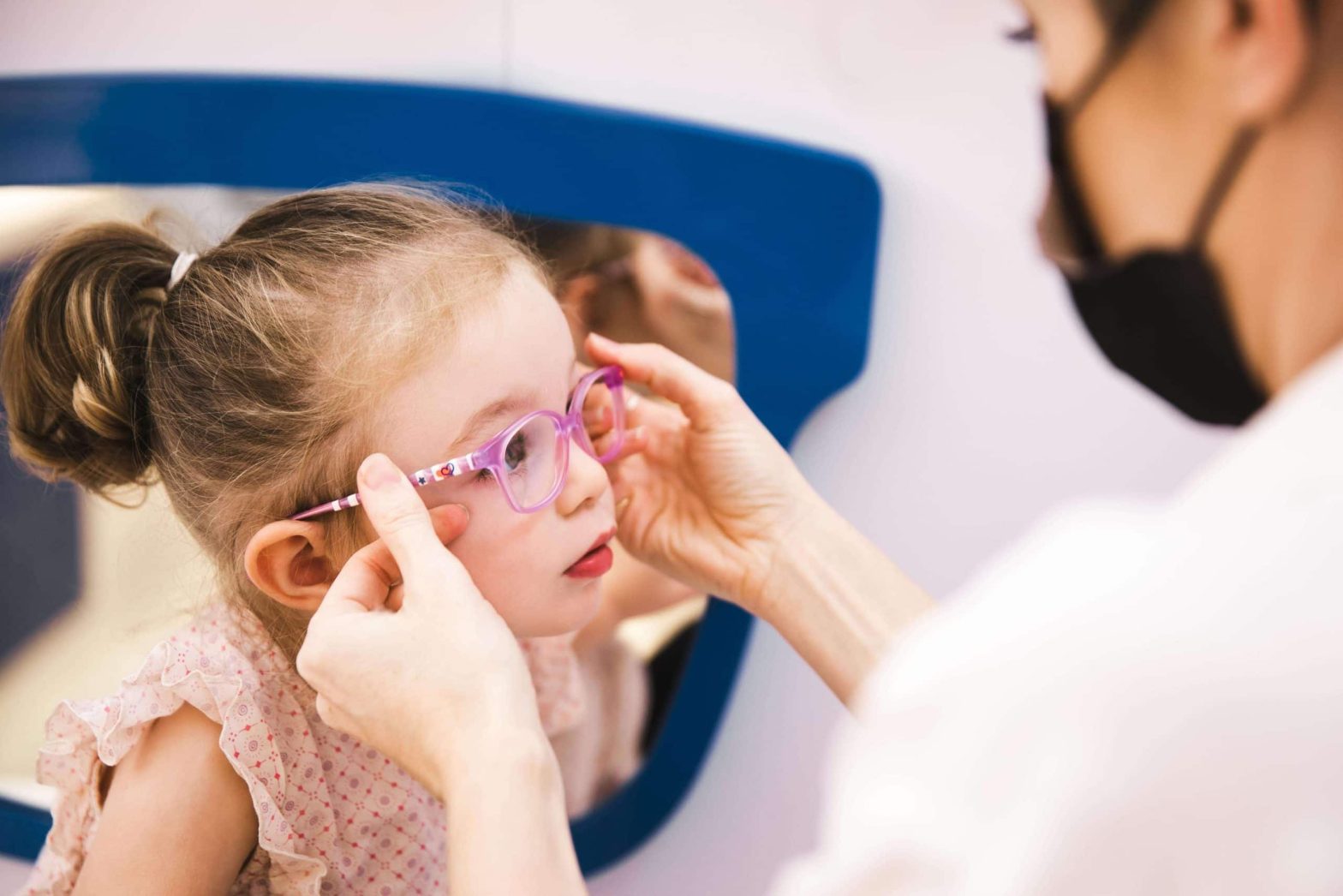
What happens during a children’s vision screening?
A paediatric eye exam is tailored to suit a child’s age and developmental level. Let us take a closer look at what happens during an exam:
- Meeting Multiple Specialists: Your child will be seen by several specialists, including orthoptists, optometrists, and paediatric ophthalmologists, ensuring a comprehensive review of their eye health.
- Vision testing: Age-appropriate methods to assess how well your child sees with our highly qualified orthoptic team.
- Eye alignment and movement: This exam includes checks to see how well both eyes work together and detect any strabismus (misalignment of the eyes).
- Eye examination: The eyes’ internal structures are examined in detail, sometimes with pupil dilation or using specialist child-friendly imaging if necessary.
What are the treatment options for Children’s eye conditions
When it comes to treating your child’s eye condition, there are both non-surgical and surgical options available to help ensure they receive the best care possible. At Moorfields Eye Hospital Dubai, we provide advanced treatments supported by the latest technology:
Non-Surgical Treatments
- Eyeglasses or contact lenses: The simplest and most common treatments for refractive errors such as myopia, hypermetropia, and astigmatism in children. Eyeglasses are often the first line of treatment to help your child see better, while contact lenses offer an alternative to glasses for active children who might find glasses cumbersome.
- Myopia management includes specialist spectacle lenses, orthokeratology (Ortho-K), eyedrops (low-concentration atropine), and soft contact lenses. We will assess your child’s eyes and work with you to find the most appropriate solution to help slow down their myopia (short-sightedness). There is a wide range of options, and at Moorfields, we have access to the latest technology.
- Patching: Used primarily to treat amblyopia (lazy eye), patching involves covering the stronger eye so the weaker eye is forced to work harder. This method can help improve vision in the affected eye over time.
- Atropine occlusion: Drops can be used in the treatment of lazy eye to blur the vision in the ‘good eye’ to encourage the brain to use the weaker eye, helping to improve the vision.
Surgical Treatments
For conditions such as squint (Strabismus), where non-surgical methods did not treat the condition, surgical intervention may be recommended. This procedure corrects misalignments in the eyes, improving not only eye alignment but also depth perception and overall vision.
Final thoughts
Children’s eye health is crucial to their overall well-being. In a vibrant city like Dubai, where academic and visual demands are high, regular eye exams with children’s eye doctor are one of the most effective ways to support your child’s learning, behaviour, and social development.
If your child has not had an eye exam recently, or if you have noticed any signs of visual difficulty, it is important to get their eyes checked with a paediatric ophthalmologist. Early detection prevents future complications and gives children the best possible start in life.
At Moorfields Eye Hospital Dubai, we are here to help your child see the world clearly—now and for years to come. Let us work together to ensure your child’s vision sets them up for success.
Frequently Asked Questions
1. Can screen time affect my child’s vision? Yes, it can. With digital screens becoming a regular part of daily routines, whether for learning or leisure, there has been a noticeable rise in eye strain and early-onset myopia in children (short-sightedness). Staring at screens for prolonged periods, especially up close, can cause discomfort, blurry vision, headaches, and fatigue. It may also encourage poor visual habits, such as holding devices too close to the eyes or forgetting to blink, which affects tear film stability and eye moisture.
To help your child, include techniques like the 20-20-20 rule (taking a 20-second break every 20 minutes to look at something 20 feet away), setting appropriate screen limits, and ensuring balanced visual activities, such as outdoor playtime. These small habits can go a long way in protecting your child’s developing vision.
2. My child is afraid of doctors—how do you handle nervous children? We understand that visiting a hospital can be daunting for little ones, especially if it is their first eye exam. That is why our entire paediatric eye care environment is designed to feel calm, safe, and welcoming. Our staff are specially trained in working with children and use gentle, playful, and engaging techniques to build trust.
We allow children to familiarise themselves with the equipment, explain each step in a fun and friendly way, and offer positive reinforcement throughout. Parents are always encouraged to stay by their child’s side during the exam, and we never rush the process. Our goal is not just to complete the assessment but to make it a positive experience your child will be comfortable returning to.
3. How long does a children’s eye exam take? A typical comprehensive children’s eye exam takes around 30 to 60 minutes. The exact duration depends on your child’s age, cooperation, and whether additional tests, such as pupil dilation or retinal imaging, are needed. We always aim for a thorough yet efficient experience, balancing accuracy with your child’s comfort.
You will have time to discuss any concerns, and we will ensure your child gets the focused attention they deserve. We recommend allowing a little extra time in your schedule, especially for first-time visits or if your child has specific vision needs.
4. Can poor vision affect my child’s behaviour or school performance? Yes, and quite significantly. Many children with vision issues may appear distracted, inattentive, or reluctant to engage in activities like reading or sports, not because of behavioural problems but simply because they are not seeing clearly. This can lead to frustration, low self-esteem, or even being misdiagnosed with learning difficulties.
A simple eye exam can reveal if vision is the underlying cause. With early intervention, whether through glasses, eye exercises, or other treatments, we can make a dramatic difference in your child’s ability to learn and thrive both academically and socially.
5. Do you offer eye care for children with special needs? Absolutely. We are proud to offer inclusive and compassionate eye care for children with developmental delays, autism, Down syndrome, and other neurological, behavioural or learning conditions. Our team takes extra time and care to understand each child’s unique needs, tailoring our approach accordingly.
Whether it means adapting communication, simplifying tests, or creating a sensory-friendly environment, we aim to make every child’s experience as smooth and positive as possible. Every child deserves clear vision; we are here to help them achieve it.
6. How often should children with glasses or existing conditions be seen? Children who already wear glasses or have known vision conditions should typically be seen every 6 to 12 months, depending on their case. Frequent monitoring allows us to check for prescription changes, ensure treatments are working effectively, and make timely adjustments.
At Moorfields Eye Hospital Dubai, we offer ongoing care and support as your child grows. Regular follow-ups are a key part of safeguarding their long-term visual development.
This blog on Oculoplastic Surgery has been contributed by Dr. Rafic Antonios, Specialist Oculoplastic Surgeon.
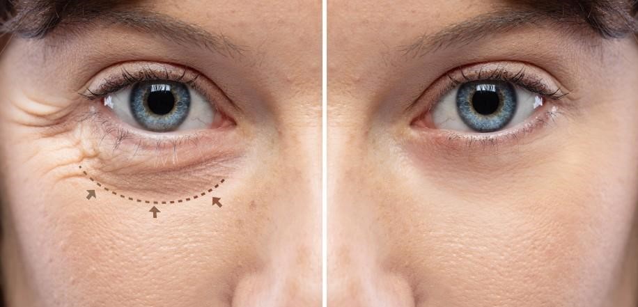
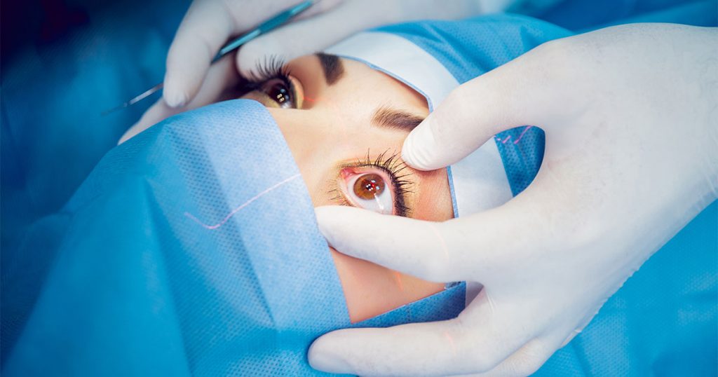
Oculoplastic surgery, often known as eyelid surgery, is a specialised field of medicine focusing on the areas around the eyes and the face. This includes the eyelids, orbit (eye socket), and lacrimal (tear) system. The purpose of these procedures ranges from aesthetic enhancements to significant functional improvements, including improvements in vision.
The Interplay Between Oculoplastic Surgery and Vision
Generally, the eyes and the surrounding structures work harmoniously to deliver clear, uninterrupted sight. However, droopy eyelids (ptosis) or eyelid malposition can disrupt this equilibrium, impairing vision. That’s where oculoplastic surgery steps in.
Addressing Common Concerns Through Surgery
Droopy eyelids, a common age-related condition, can obstruct the field of vision, making daily activities like reading or driving challenging. Oculoplastic surgery corrects this by tightening the muscles that lift the eyelid, thereby widening the visual field. Similarly, conditions like entropion (inward-turning eyelid) or ectropion (outward-turning eyelid) can cause discomfort, tear overflow, and blurred vision. Surgery to reposition the eyelid reinstates normalcy, relieving the symptoms and restoring vision clarity.
The Transformative Potential of Oculoplastic Surgery
In conclusion, oculoplastic surgery is not just about cosmetic enhancement. It’s a field aimed at restoring or maintaining the eye’s optimal functionality, significantly impacting vision. While the thought of surgery can seem daunting, understanding its potential benefits can provide reassurance. Consulting with an oculoplastic surgeon could be a transformative step for your vision if you are experiencing vision disruption due to eyelid or surrounding orbital structure issues.
If you are considering oculoplastic surgery in Dubai or want to learn more about how these procedures can enhance your vision and appearance, we are ready to help you by providing personalised care tailored to your unique needs and goals.

Orthoptist Sehar Mann has contributed to this blog on Stages of visual development in Children.
We are all born with blurry vision, and our vision develops in the first 7-8 years of life but is most important when we are babies and toddlers.
Understanding the stages of visual development in babies and toddlers and how they perceive colours can help you provide the correct visual stimulation in their early months of life.
Stages of Visual Development
- The Newborn Stage (0-2 months): Newborn babies can only see objects about 8 to 12 inches from their face – roughly the distance between your face and theirs during feeding or cuddling. They are drawn to high-contrast patterns, like black and white since their eyes are still developing.
- The Infancy Stage (2-4 months): During this period, your baby’s vision starts to improve gradually. They follow moving objects and show interest in colourful toys and pictures. It is normal for their eyes to cross or wander as their muscles strengthen occasionally.
- The Exploratory stage (4-6 months): At 4 to 6 months, babies reach for objects and have better depth perception as they can see more colours more clearly. This is an ideal time to introduce more colourful toys and books with large, bold pictures to stimulate their developing vision.
How Toddlers Perceive Colours
Understanding how toddlers perceive colours can help you create an engaging and stimulating environment for them. Here are some tips to help them:
- High Contrast is Key: In the early months, your baby is drawn to high-contrast colours, such as black and white because they are easier for their developing eyes to distinguish. Consider incorporating these colours into your baby’s nursery decorations or toys.
- The world of Pastels: As babies grow, they will notice more subtle colours like pastels. Soft blues, pinks, and yellows can be soothing and visually appealing for them.
- Vibrant Colours: Babies can see a broader range of colours in the toddler stage. Bright and vibrant colours like red, green, and blue can capture their attention and stimulate their visual development.
Visual Stimulation in the First 6 Months
Visual stimulation is essential for development during the first six months. Here are some simple ways to provide the correct visual experiences:
- Face-to-Face Interaction: Spending quality time making eye contact and talking to your baby can help develop their vision. They love to gaze at your face, and this helps strengthen their visual focus.
- Colourful mobile Toys: Hang a colourful mobile above the crib. The gentle movement and bright colours will intrigue your baby and encourage them to track the objects.
- Picture Books: Choose picture books with large, bold images and vibrant colours. Reading to your baby fosters bonding and enhances their visual recognition skills.
- Tummy Time: Give your baby plenty of tummy time. This helps them develop strong neck and shoulder muscles, which are essential for visual exploration.
- Mirrors: Babies are often fascinated by their reflection. A baby-safe mirror can provide endless entertainment and help with self-recognition.
Watching your toddler’s visual development is an exciting journey that requires your support and engagement. By understanding the stages of visual development and how your baby perceives colours, you can create a stimulating environment that encourages healthy growth and curiosity. Remember, every baby is unique, so enjoy the discovery process together and celebrate each new milestone.
At Moorfields Eye Hospital Dubai, our dedicated paediatric department is staffed with expert ophthalmologists, optometrists, and orthoptists, all passionately dedicated to the eye care needs of children.

Strabismus, commonly called ‘crossed eyes,’ impacts approximately 2% – 4% of children worldwide. This condition results in the misalignment of the eyes, causing them to point in different directions and disrupting binocular vision.
Strabismus is primarily seen in infants and young children and is not just a cosmetic concern. It can significantly impact vision development and depth perception, essential for numerous activities, from catching a ball to crossing a street. When the eyes do not align properly, the brain may start to ignore the image from the eye that is not aligned to prevent seeing double, causing ‘amblyopia,’ also known as a lazy eye.
Emphasising early detection is crucial with strabismus, as the initial years of a child’s life are vital for visual development. Any signs of the condition, such as squinting, tilting the head while looking at objects, or noticeable misalignment of the eyes, should prompt an immediate medical consultation.
The treatment journey for a child with strabismus often involves a team of specialists. An optometrist performs regular eye exams and prescribes glasses if necessary. An orthoptist specialising in strabismus and amblyopia devises personalised eye exercises to improve eye coordination. Lastly, a paediatric eye consultant oversees the child’s treatment, deciding when surgical intervention is necessary and ensuring the various treatments align to maximise visual development and overall quality of life.
In essence, strabismus affects not only the physical appearance but also the functional vision of a child. Therefore, recognising and addressing strabismus in its early stages is crucial for optimal eye health and developmental growth.
If you notice any of the signs or symptoms of Strabismus, please seek care from a specialised ophthalmologist to help your child’s vision develop correctly.
At Moorfields Eye Hospital Dubai, our dedicated paediatric department is staffed with expert ophthalmologists, optometrists, and orthoptists, all passionately dedicated to the eye care needs of children.
This blog on Selective Laser Trabeculoplasty for glaucoma treatment was authored by Dr. Salman Waqar, a Consultant Ophthalmologist in Cataract and Glaucoma Surgery.
Living with glaucoma often means meticulous eye drop management and close intraocular pressure (IOP) monitoring. Thankfully, advancements in eye care, such as Selective Laser Trabeculoplasty (SLT), offer a more refined to manage this condition.
What is SLT?
Selective Laser Trabeculoplasty is a minimally invasive procedure designed to help control glaucoma by decreasing IOP. One of its standout features is the potential to lessen, or in some cases, even eliminate, the dependence on glaucoma eye drops, especially beneficial for those with open-angle glaucoma.
How Does It Work?
Imagine the eye’s drainage system, the trabecular meshwork, as a sink’s drain. Over time, this drain can become less efficient, leading to increased IOP. During this procedure, a precision-targeted laser is applied to this drainage system. Unlike other methods, Selective Laser Trabeculoplasty ensures no thermal damage to the nearby tissue. The treatment encourages the drainage system to work more efficiently by promoting beneficial biological changes within its cells. As the drainage improves, the IOP naturally decreases.
Why Consider SLT?
Selective Laser Trabeculoplasty is revolutionary in its approach. Unlike more aggressive surgeries, it targets only specific cells, ensuring surrounding tissues remain untouched. This selectivity means a less invasive procedure, minimal risks, and quicker recovery times.
However, it’s essential to remember that while it is highly effective, it might not completely negate the need for glaucoma eye drops for all patients. For some, it can notably decrease the amount or frequency of medication, leading to improved comfort and better adherence to treatment.
In Conclusion
Selective Laser Trabeculoplasty is a promising option in glaucoma care. Potentially reducing our reliance on daily eye drops offers a more convenient yet effective way to keep glaucoma in check and safeguard our vision. If you are considering this procedure, discussing it with a glaucoma specialist is recommended to tailor the best care for your eyes.
Request an Appointment