Posterior Vitreous Detachment

PVD is a common degenerative change, which affects one or both eyes in many people after middle age. It may present earlier in shortsighted patients or those who have sustained traumas to the eyes.
Thickening of the jelly casts shadows on the retina and are seen as floating shapes. These black “floaters”in your vision move with the eye and then settle as the eye rests. These are often described by patients as a “cobweb” or “insects”.
You may also be aware of flashing lights, like little flickers in the outer periphery. Usually these do not highlight a problem, however, it is important to have the eye thoroughly checked, as occasionally a retinal tear or a retinal detachment may occur.

Post-Operative Instructions

Following Retinal Surgery on leaving the hospital you are advised to have a quiet evening at home and to avoid strenuous exercise.
For General Anaesthetic patients, as above and:

  • Do not drive a vehicle
  • Do not make any crucial financial decision
  • Do not eat heavy meals or drink alcohol for 24 hours after being discharged

Paediatric Strabismus

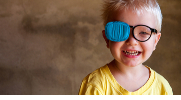
Facts

A squint is a condition where your eyes look in different directions. One eye turns inwards, outwards, upwards or downwards while the other eye looks forwards. The medical name for a squint is strabismus.

The misalignment of the eyes can be caused by different factors. It can be an early developmental problem where the brain struggles to identify that the two eyes should work as a pair. It can be caused by an abnormality with the eye muscles or an uncorrected vision problem, such as myopia (shortsighted), hypermetropia (longsighted) or Astigmatism.

When to see a doctor?

Squints in children are relatively common. They usually develop before a child is five years of age, but they can appear later.

Up to around three months of age, many babies occasionally squint as their vision develops. This is normal and nothing to worry about. If your child still has a squint after this age, you should visit your Doctor. It is very important that a squint is picked up and treated as early as possible to avoid vision problems developing. If a squint is identified when a child is young, there is a good chance that it will be successfully treated.

Can adults get a squint?

Occasionally, squints that have been corrected during childhood reappear in adulthood. New squints in adults, without any history of a squint in childhood, can be caused by problems with the ocular muscles and/or the eye movement system. You should visit your Doctor as soon as possible if you develop a new squint. They should refer you to an ophthalmologist who will carry out an examination to identify the cause.

Squints that affect adults may cause double vision because the brain has been trained to collect images from both eyes. Squints may also cause a cosmetic problem in adults; in such cases, the appearance of a squint can lead to low self-esteem

What is Amblyopia?

Amblyopia is also known as a ‘lazy eye’. Amblyopia is an early childhood condition where a child’s eyesight in one eye does not develop as it should. The problem is usually in just one eye, but can sometimes affect both of them. Amblyopia affects approximately 2% of children.

When a patient has amblyopia the brain focuses on one eye more than the other, virtually ignoring the ‘lazy eye’. If that eye is not stimulated properly the visual brain cells do not mature normally.

What causes a ‘lazy eye’?

Anything that interferes with clear vision in either eye during the critical period (birth to 6 years of age) can cause amblyopia. The most common causes of amblyopia are constant strabismus (constant turn of one eye), anisometropia (different vision/prescriptions in each eye), and/or obstruction of an eye due to cataract, trauma, lid droop, etc.

Why does my child need to wear a patch?

Occlusion (patching) is used to make a lazy eye work on its own and so improve the vision by encouraging the development of the nerve pathways from that eye to the brain. The patch is worn over the good eye and the amount of time the patch must be worn is decided by the Orthoptist/Ophthalmologist and relates to the extent of the visual problem. If patching is implemented early on, a good level of vision can be achieved. When patching is started in an older child, it is more difficult to achieve good vision.

What is an Orthoptist?

An Orthoptist specialises in diagnosing and treating visual problems involving eye movement and alignment.

The Orthoptist at Moorfields Dubai provides clinical support to all the specialist services at the hospital. She sees both adults and children who have strabismus (a squint), disorders of eye movements, or binocular vision.

What is an Optician?

An Optician will see adults and children for refraction; with this assessment, an optometrist can determine the optical power of the eye, the presence of any “refractive” error that requires spectacle correction, and the best vision that an eye can achieve with an appropriate correction. Younger children have drops to make the pupil (the dark center of the eye) larger and this makes the test more accurate.

What is an Ophthalmologist?

An Ophthalmologist is a specialist in medical and surgical eye problems. Since ophthalmologists perform operations on eyes, they are considered to be both surgical and medical specialists. They will check both the structure and health of the eye. They will make the final decision on the management and will do any surgical procedures required.

Myopia

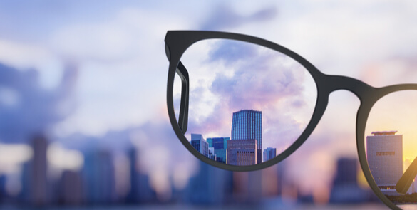
Myopia is a common refractive condition which causes individuals to be near-sighted: they see near objects clearly but distant objects are blurry. Myopia occurs when the cornea and lens focus the light in front of the retina instead of exactly on it. Symptoms of myopia include; difficulty seeing distant objects, squinting frequently, holding books or other objects very close to the face, difficulty seeing writing on signs or watching television and difficulty with driving (particularly at night). Myopia should be diagnosed by a qualified Optometrist, Ophthalmic Surgeon or Eye Specialist. Myopia is best treated with eyeglasses or contact lenses which compensate for the elongated shape of the eye allowing the light to focus properly on the retina. Refractive surgery is another option that eliminates dependence on glasses or contact lenses.

Lacrimal Probing in Children

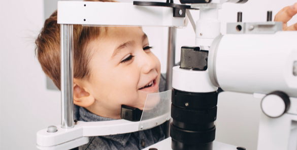
The tear duct is a channel/passage which runs from a tiny opening in the medial lids through the bone to the inside of the nose, and drains the tears and mucus the eye produces. It should open just before or just after birth but sometimes remains blocked for a considerable time after that, causing watering and discharge from the eye. It is harmless, and does not affect the health of the eye or the vision, although it can make the eyelids red and sore and slightly increases the frequency of infective conjunctivitis.

Keratoconus

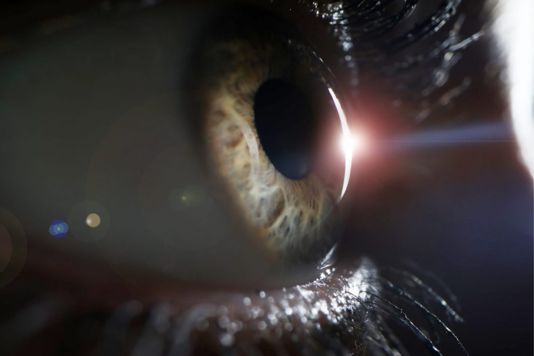
Keratoconus is a progressive thinning of the cornea. The cornea is the clear front window of the eye, which, along with the intra-ocular lens, focuses light on to the retina. The cornea normally is a smooth, round dome-shaped structure; however in keratoconus it becomes very thin and irregular and it starts to protrude from the centre or below the centre like a cone. This causes blurry vision that is often not completely correctable with glasses.
The disease is multifactorial in origin but there is a strong genetic component which makes it more frequent in certain parts of the world or within certain families.

Intravitreal Injection

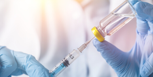
The macula is the central part of the retina at the back of the eye. It is responsible for fine vision (reading, writing, watching television, and recognising faces). Patients with diabetes may develop macular oedema (swelling of the retina) due to leaking of fluid from blood vessels. This causes the vision to become blurred.
A course of three injections is recommended with each injection administered one month apart. The procedure is carried out in a clean environment using sterile technique. The eye is cleaned and local anaesthetic drops are given to numb the eye.
The eye may or may not be covered after the injection. If a pad is applied, this may be removed when you reach home.

Hypermetropia

A refractive condition of the eye in which vision is better for distant objects than for near objects. It can be called far sightedness or hypermetropia. Symptoms of Hypermetropia can include; blurred vision, asthenopia (eye strain), accommodative dysfunction, binocular dysfunction, amblyopia and strabismus. It results from the eyeball being smaller than average, causing images to be focused behind the retina. Hypermetropia should be diagnosed by a qualified Optometrist, Ophthalmic Surgeon or Eye Specialist.
A full Optometric Examination should be performed to assess the degree and extent of the problem. Eyeglasses and contact lenses are the treatment of choice for most people with far sightedness but refractive surgery can also cure some cases of hyperopia.

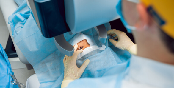
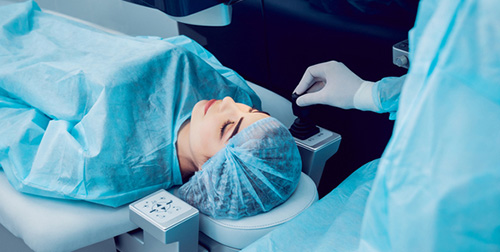
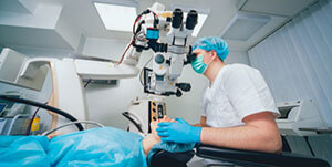
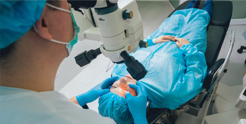
High Precision Refractive Surgery

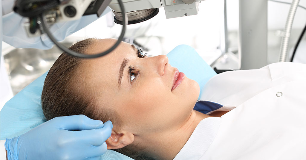
When you decide on an eye laser treatment, you expect the best possible results. The more fully developed the methods are, the better the outcome will be. The SCHWIND AMARIS 750S offers you the leading technology for your laser treatment – superior in all important aspects: Speed, precision, safety and comfort.

Diabetic Retinopathy

Introduction

  • Diabetic retinopathy is a complication of diabetes and leads to high blood sugar, resulting in retinal disease, which can interfere with its ability to transmit images to the brain through the optic nerve.
  • Blood vessels in the retina play an important role in supplying it with oxygen and nutrients, which keep it healthy and working effectively.

Diabetic Retinopathy can result in damage to the blood vessels; these may then bleed, leak or become blocked leading to cell damage in the retina itself.
There are varying forms and levels of severity of diabetic retinopathy – for example, when the retina becomes very damaged, new blood vessels may grow on it and can burst, leading to bleeding and blurred vision. If the macula (the central area of the retina) is affected – this is called Diabetic Maculopathy – the disease has reached a much more advanced and serious stage.
High blood pressure combined with diabetes leads to an even more dangerous condition.

Types of Diabetic Retinopathy

Non Proliferative Diabetic Retinopathy
Generally, this type of diabetic retinopathy does not affect vision because at the initial stage there are just a few enlarged blood vessels, with very minimal bleeding and leaking in the retina.
An examination of the retina by an ophthalmologist will reveal some marks indicating the presence of the condition.
Proliferative Diabetic Retinopathy
This condition can lead to seriously impaired vision as blood vessels grow in the retina and the threat is bleeding from these vessels which can lead to retinal damage, and even to retinal detachment at the back of the eye. Laser treatment is essential to avoid serious long term damage.
Diabetic Maculopathy
Diabetic maculopathy occurs when blood vessels leak into the central area of the retina, which can lead to it swelling and affecting the quality of vision. Laser procedures, injections or surgery are the main treatment options.
Exudates are deposits in the retina from leaky vessels. Haemorrhages in the retina of varying sizes.
Microaneurysms.
Optic Nerve with abnormal new vessels that have bled. Scars from previous laser treatment.
Vitreous haemorrhage in front of the retina.
End stage proliferative Diabetic Retinopathy with fibrousbands.
Retinal Detachment.
Cross sectional scan showing thickening of the macula and exudates.
Exudates (yellow) and haemorrhages in the central retina

Can it be prevented?

Compliance with treatment for diabetes helps control blood sugar levels and blood pressure and so the serious complications of diabetes, including diabetic retinopathy.
Treatment compliance along with regular check ups to monitor blood pressure will help avoid the serious long term effects of diabetes; positive lifestyle choices from balanced diet to regular exercise and not smoking all have a beneficial effect.

What can I do?

Early diagnosis and treatment of diabetes and diabetic retinopathy will generally help prevent serious vision loss for most patients and regular visits to the doctor and ophthalmologist are an essential part of the monitoring process.

Corneal Transplantation (PK)

Why do you need a corneal transplant?

The cornea is a window of transparent tissue at the front of the eyeball. It allows light to pass into the eye and provides focus so that images can be seen. Various diseases or injury can make the cornea either cloudy or out of shape. This prevents the normal passage of light and affects vision.
A cloudy cornea can be replaced by a healthy one from a donor to restore vision.If the full thickness of the cornea is affected by disease, then a full thickness transplant is performed. This is known as a penetrating keratoplasty.

Benefits of penetrating keratoplasty

Improved vision

  • Approximately 75% of transplant recipients have vision sufficient to drive legally but may need glasses or contact lenses or sometimes further surgery for best results.
  • It may take up to 18 months until the full improvement in vision is appreciated.

Risks of penetrating keratoplasty

Rare but serious complications

  • Sight-threatening infection (1 in 1,000)
  • Severe haemorrhage causing loss of vision
  • Retinal detachment
  • Severe inflammation or other rare causes of loss of vision

Corneal transplant rejection
A corneal transplant can be identified and attacked by your immune system. This happens in one in six patients in the first two years after transplantation and can cause graft failure. It can often be reversed if anti-rejection medication is started promptly.
Rejection remains a possibility for your lifetime.
Graft failure
When a graft fails, your cornea becomes cloudy again and your vision becomes blurred. This happens in one in 10 transplants for keratoconus in the first 10 years.
Glaucoma
This can usually be controlled by eye drops, but occasionally requires surgery and can damage the sight.
Cataract
This can be removed surgically.

About the operation

The operation
The operation is performed under general or local anaesthetic. The operation takes about one hour. A central 8mm button of your cornea is removed and a similar-sized button of the donor cornea is stitched in with tiny stitches (see front cover). These cannot be felt or seen. The abnormal cornea, which is removed, is sent to our pathology laboratory for examination under a microscope.
After the operation
You will usually be examined by the surgical team after the surgery and can generally go home the same day. You will be seen again within one week in the outpatient clinic and regularly thereafter (approximately six visits in the first year). We generally recommend that you take two weeks off work – discuss your individual circumstances with your doctor. You will need to use anti-rejection eye drops for at least six months and in some cases indefinitely. Individual
stitches may be removed from three months after the operation, but complete stitch removal is not performed until at least one year after the procedure.
What if my transplant fails?
A failed transplant can be replaced in a procedure known as a regraft, but the risk of subsequent rejection and failure increases each time for regrafts.
The percentages of full-thickness corneal grafts that are still functioning well five years after the operation under various conditions are:

Condition %
Keratoconus 95
Fuchs’ dystrophy 80-90
Stormal Scar 80-90
Stromal dystrophies 80-90
Bullous keratopathy 50-80
Bacterial infections 50-80
Herpetic keratitis 50-80
Fungal infection 0-50
3rd or higher number regraft 0-50
4 quadrants of blood vessels 0-50
Inflammation at time of surgery 0-50
Severe ocular surface disease 0-50
Grafts greater than 10mm 0-50

Consenting for information sharing
We are required to share your information with the Eye Bank who supply donor corneas, to ensure high quality transplant material.
Corneal transplant rejection
Rejection needs urgent treatment as this can lead to failure of the transplant and loss of vision.
Symptoms of rejection are:

  • Red eye
  • Sensitivity to light
  • Visual loss
  • Pain

If you experience any of these symptoms, you should immediately call our 24-hour emergency phone line 055 516 1586.

Corneal Transplantation (EK)

Why do you need a corneal transplant?

The cornea is a window of transparent tissue at the front of the eyeball. It allows light to pass into the eye and provides focus so that images can be seen. Various diseases or injury can make the cornea either cloudy or out of shape. This prevents the normal passage of light and affects vision.
The cornea has three layers (thin outer and inner layers and a thick middle layer). In some diseases, only the inside layer (endothelium) is affected, causing corneal oedema (swelling) and clouding (see below). Endothelial keratoplasty is a modern technique to replace the inside layer of your cornea with the inside layer from a donor cornea through a relatively small incision (opening).

Benefits of endothelial Keratoplasty

Improved vision
The majority of transplant recipients have sufficiently good vision to be able to drive legally although many need glasses. It can take up to six months until the full improvement is appreciated. Comfort is improved in some cases.

Risks of endothelial Keratoplasty

Rare but serious complications

  • Sight-threatening infection (1 in 1,000)
  • Severe haemorrhage causing loss of vision
  • Retinal detachment
  • Severe inflammation or other rare causes of loss of vision

Corneal transplant rejection
A corneal transplant can be identified and attacked by your immune system. This happens in between 6% and 10% of DSAEK recipients in the first two years after transplantation and can cause graft failure. It can often be reversed if anti-rejection medication is started promptly. Rejection remains a possibility for your lifetime. The rejection risk in DMEK appears to be lower than in DSAEK.
Graft failure
When a graft fails, the cornea becomes cloudy again and vision becomes blurred.
Glaucoma
This can usually be controlled by eye drops, but occasionally requires surgery and can damage your sight.
Graft dislocation
About 10% of endothelial grafts dislocate and need to be repositioned in theatre.
Cataract
This can be removed surgically.

Possible advantages of EK over full-thickness graft

  • Faster recovery
  • Fewer stitches, which means that the shape of the cornea is more “normal” and you are less dependent on glasses/ contact lens
  • Smaller wound so fewer wound complications such as leakage or wound rupture after accidental injury

About the operation

The operation
The operation is usually performed under local anaesthetic and takes about one hour. Through a small incision (opening), your endothelium is removed and an 8.5mm disc of donor endothelium is inserted and pressed in position against the back of your cornea by a bubble of air. You will need to lie flat for one hour after the operation. Usually, only two stitches are used to close the incision.
After the operation
You will usually be examined by the surgical team after your surgery and can generally go home the same day. You will be seen again the next day and within one week to make sure the graft stays in position. You will have about six visits to the outpatient clinic in the first year. We generally recommend that you take two weeks off work – discuss your individual circumstances with your doctor. You will need to use anti-rejection eyedrops for at least six months and in some cases indefinitely. The stitches are usually removed at about three months.

What if my transplant fails?

A failed transplant can be replaced in a procedure known as a regraft. However, the risk of subsequent rejection and failure increases each time for second and subsequent regrafts.
Consenting for information sharing
We are required to share your information with the Eye Bank who supply donor corneas, to ensure high quality transplant material.
Corneal transplant rejection
Rejection needs urgent treatment as this can lead to failure of the transplant and loss of vision.
Symptoms of rejection are:

  • Red eye
  • Sensitivity to light
  • Visual loss
  • Pain

If you experience any of these symptoms, you should immediately call our 24-hour emergency phone line 055 516 1586.