MBBS, MS (Ophth), MRCOphth, FRCOphth Consultant Ophthalmologist Vitreo-Retinal Surgeon GCAA Approved Specialist Medical Examiner]Dr. Muralidharan Upendran is a Consultant Ophthalmologist with sub-specialist training in Vitreo-Retinal surgery. He specialises in the surgical management of retinal disorders including retinal detachment, macular disease, diabetic retinopathy and ocular trauma. He also specializes in the medical management of retinal vascular… Continue reading Dr. Muralidharan Upendran
Dr. Muralidharan Upendran
MBBS, MS (Ophth), MRCOphth, FRCOphth Consultant Ophthalmologist Vitreo-Retinal Surgeon GCAA Approved Specialist Medical Examiner]Dr. Muralidharan Upendran is a Consultant Ophthalmologist with sub-specialist training in Vitreo-Retinal surgery. He specialises in the surgical management of retinal disorders including retinal detachment, macular disease, diabetic retinopathy and ocular trauma. He also specializes in the medical management of retinal vascular… Continue reading Dr. Muralidharan Upendran
Dr. Osama Giledi
MBBch, FRCSEd Consultant Ophthalmologist Specialist in Cataract, Cornea and Refractive Vision Correction Surgery GCAA Approved Specialist Aeromedical Medical Examiner Associate Professor of Ophthalmology (Adjunct) Dr Osama Giledi is a highly experienced consultant ophthalmologist who specialises in Cornea, Anterior Segment, Cataract and Refractive Surgery. He is also skilled in managing ocular surface problems including severe dry… Continue reading Dr. Osama Giledi
Types of Diabetic Retinopathy

Introduction Diabetic retinopathy is a complication of diabetes and leads to high blood sugar, resulting in retinal disease, which can interfere with its ability to transmit images to the brain through the optic nerve. Blood vessels in the retina play an important role in supplying it with oxygen and nutrients, which keep it healthy and… Continue reading Types of Diabetic Retinopathy
Corneal Transplantation (PK)
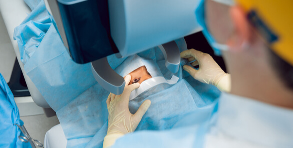
Why do you need a corneal transplant? The cornea is a window of transparent tissue at the front of the eyeball. It allows light to pass into the eye and provides focus so that images can be seen. Various diseases or injury can make the cornea either cloudy or out of shape. This prevents the… Continue reading Corneal Transplantation (PK)
Corneal Transplantation (EK)
Why do you need a corneal transplant? The cornea is a window of transparent tissue at the front of the eyeball. It allows light to pass into the eye and provides focus so that images can be seen. Various diseases or injury can make the cornea either cloudy or out of shape. This prevents the normal passage of light and… Continue reading Corneal Transplantation (EK)
Corneal Transplantation (DALK)
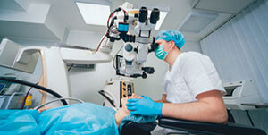
Why do you need a corneal transplant? The cornea is a window of transparent tissue at the front of the eyeball. It allows light to pass into the eye and provides focus so that images can be seen. Various diseases or injury can make the cornea either cloudy or out of shape. This prevents the… Continue reading Corneal Transplantation (DALK)
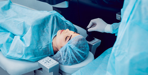
Cataract Surgery Treatment
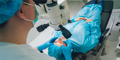
Facts The term cataract derives from the view we get when looking through a waterfall. A cataract is the clouding or opacity of the lens inside the eye. The lens has the shape of a lentil and lies behind the coloured part of the eye, the iris. In a normal eye, this lens is clear.… Continue reading Cataract Surgery Treatment
Blepharitis
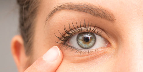
Facts Common condition that causes inflammation of the eye lids and can affect people of all ages. It usually affects the rim of the eyelids of both eyes and usually is not serious but still irritating and uncomfortable. Eyelids may become crusty and scaly and eyes may feel gritty and very tired with increasing irritation… Continue reading Blepharitis
Atropine Drops
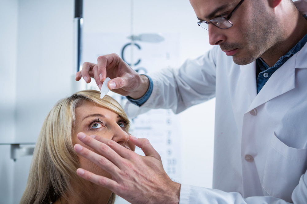
Atropine Eye Drops: Uses, Benefits & What to Know Atropine has two effects when instilled in the eyes: Dilates the pupil (makes the black part of the eye larger). Stops the eye from focusing properly – blurring the child’s near vision temporarily. Why has my child been prescribed Atropine? Atropine has been prescribed to enable… Continue reading Atropine Drops
Astigmatism information

Facts Astigmatism is a treatable eye condition that can cause blurred vision and headaches. It is a refractive condition in which the eye’s optical system is incapable of forming a point image for a point object (images are misconstrued). The refractive error of the astigmatic eye stems from a different degree of refraction in different… Continue reading Astigmatism information