


About Retina
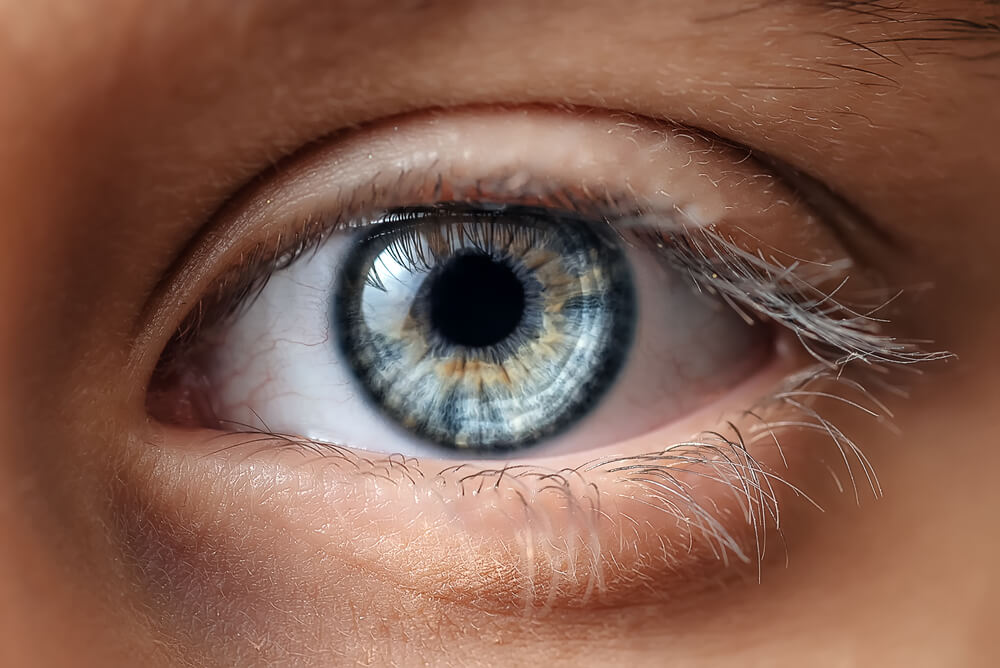
The retina is a vital component of the eye that plays a crucial role in our vision. As a thin layer of tissue near the optic nerve at the back of the eye, the retina acts as a powerful sensory receptor. Its role is to convert the light focused on it by the eye’s lens into electrical signals, then transmitted to the brain.
The retina allows us to perceive colours, differentiate between light intensities, and perform essential daily tasks such as reading and driving. The retina’s photoreceptor cells and optic nerve communicate visual information to the brain.
However, when the retina is damaged, it can lead to vision loss and even blindness. Therefore, addressing retinal conditions to protect and preserve your eyesight quickly is crucial.
Our retina specialists are experts in diagnosing and treating various retinal conditions. Some of the commonly encountered retinal conditions include:
- Age-related macular degeneration: This condition primarily affects older adults and can cause central vision loss, impairing activities like reading and recognising faces.
- Retinal detachment: A severe condition where the retina separates from the back of the eye, often resulting from trauma or other underlying eye disorders. Immediate medical attention is necessary to prevent permanent vision loss.
- Diabetic-related eye conditions: Individuals with diabetes are at risk of developing retinopathy, a condition that damages the blood vessels in the retina. Diabetic retinopathy can lead to vision impairment if left untreated.
- Uveitis: This inflammatory condition affects the eye’s middle layer, including the retina. Prompt treatment with a Uveitis specialist is crucial to prevent potential vision loss and complications.
- Macular hole: A macular hole is a small break in the macula, the central part of the retina responsible for sharp, detailed vision. Surgical intervention may be necessary to restore vision in severe cases.
- Floaters: Floaters are specks or cobweb-like shapes that appear to drift across your field of vision. While they are usually harmless, sudden changes in the number or size of floaters may indicate a retinal issue that requires attention.
If you experience any symptoms, such as sudden vision changes, flashes of light, or the appearance of new floaters, it is crucial to consult a retina specialist. Early detection and appropriate treatment can significantly improve outcomes for retinal conditions.
At Moorfields Eye Hospitals, our Retina and Uveitis specialists in Dubai and Abu Dhabi employ state-of-the-art diagnostic tools and advanced treatment techniques to preserve and restore your precious eyesight.
Comprehensive Assessments
We offer a comprehensive range of eye care assessments, diagnostics, surgical and non-surgical treatment services. Assessment services: (evaluation examinations may differ depending on outcome of consultation) (expandable links below)
- General Health Assessment
- Vision screening
- Color Fundus Photos of the Retina
- OCT of the Macula
- Ultrasound
- UBM
- Electrophysiology (ERG)
- Consultation with Retina Consultant
- General Health Assessment
- Vision screening
- Optos wide Field color photos
- Optos wide fields RF & IR
- Ultrasound
- UBM
- Electrophysiology (ERG)
- Consultation with Retina Consultant
- General Health Assessment
- Vision screening
- Color Fundus photos (Heidelberg)
- IR & Auto-fluorescence (AF) Fundus photos (Heidelberg)
- OCT scan
- OCTA
- Ultrasound
- UBM
- Electrophysiology (ERG)
- Consultation with Retina Consultant
Conditions & Treatment
Intravitreal Injection
About Intravitreal Injection
The macula is the central part of the retina at the back of the eye. It is responsible for fine vision (reading, writing, watching television, and recognising faces). Patients with diabetes may develop macular oedema (swelling of the retina) due to leaking of fluid from blood vessels which can result in the vision becoming blurred.
Diabetic macular oedema
Diabetic eye disease is a leading cause of blindness. It is caused by changes to the tiny blood vessels of the retina (the light sensitive layer at the back of the eye). In diabetic macular oedema, blood vessels leak fluid into the retina.
Macular Hole Surgery
About Macular Hole Surgery
If you think of your eye as a camera, the retina is like the photographic film. It is a very thin layer of tissue, which is sensitive to the image focused on it, sending the information to the brain.
At the very centre of the retina is the macula. This is a very special area of the retina, which we use for reading and recognising complex shapes. Sometimes, a hole forms in the macula, which prevents it from working normally. This affects your vision, particularly for reading and other visually demanding tasks, but it does not cause total blindness.
Posterior Vitreous Detachment
About Posterior Vitreous Detachment
Posterior Vitreous Detachment (PVD) is a common degenerative change, which affects one or both eyes in many people after middle age. It may present earlier in shortsighted patients or those who have sustained traumas to the eyes.
Thickening of the jelly casts shadows on the retina and are seen as floating shapes. These black “floaters “in your vision move with the eye and then settle as the eye rests. These are often described by patients as a “cobweb” or “insects”.
You may also be aware of flashing lights, like little flickers in the outer periphery. Usually these do not highlight a problem, however, it is important to have the eye thoroughly checked, as occasionally a retinal tear or a retinal detachment may occur.

Retinal Detachment Surgery
About Retinal Detachment Surgery
Retinal detachment is a condition when the thin lining at the back of the eye (the retina) begins to come away and separate itself from the underlying wall of the eye which contains blood vessels that supply it with vital oxygen and nutrients.
If not treated promptly, retinal detachment will lead to blindness in the affected eye.
A retinal detachment is usually caused by a tear in the retina and this is termed a Rhegmatogenous retinal detachment. There are other types of retinal detachment namely Traction retinal detachment which is usually seen in advanced diabetic retinopathy and Exudative retinal detachment (usually seen in people with inflammation). It is the Rhegmatogenous retinal detachment which needs urgent surgery in most cases.
Retinal Photodynamic Therapy (PDT)
About Retinal Photodynamic Therapy (PDT)
Photodynamic therapy (PDT) is a treatment that uses a combination of a “cold” laser and a special light-sensitive dye (Verteporfin). This is injected into the blood stream to target abnormal leaking blood vessels in the retina (nerve tissue lining at the back of the eye which detects light and allows us to see) or the layer below the retina (choroid). PDT is used in the treatment of some specific forms of wet macular degeneration and a disease called ‘Central Serous Retinopathy’ (CSR).
Retinal Vein Occlusion
About Retinal Vein Occlusion
Blocking of the retinal vein which reduces the vision is known as Retinal Vein Occlusion. This results in the accumulation of blood (retinal hemorrhages) and fluid (macular edema) in the retina and leads to a drop in the visual acuity (clarity of vision).
Diabetic Retinopathy
What is Diabetic Retinopathy?
Diabetic retinopathy is a serious complication of long-term uncontrolled diabetes resulting from persistently high blood sugar levels over the years. This condition affects the retina, the light-sensitive tissue at the back of the eye, impairing its ability to transmit clear images to the brain through the optic nerve.
At Moorfields Eye Hospitals, our retina specialists in Dubai and Abu Dhabi have extensive sub-speciality training in the diagnosis and treatment of diabetic retinopathy. Using the latest retinal imaging and advanced therapeutic techniques, they provide tailored care to slow disease progression, protect sight, and support patients in maintaining their long-term eye health and quality of life.
Diabetic Maculopathy
About
Diabetic maculopathy is a condition that occurs when the macula, the part of the eye which provides us with our central vision is damaged. The macula is responsible for fine vision (reading, writing, watching television, and recognising faces). Patients with diabetes may develop macular oedema (swelling of the retina) due to leaking of fluid from blood vessels. This causes the vision to become blurred.Diabetic Maculopathy
About
Diabetic maculopathy is a condition that occurs when the macula, the part of the eye which provides us with our central vision is damaged. The macula is responsible for fine vision (reading, writing, watching television, and recognising faces). Patients with diabetes may develop macular oedema (swelling of the retina) due to leaking of fluid from blood vessels. This causes the vision to become blurred.Age-related Macular Degeneration (AMD)
About Age-related Macular Degeneration (AMD)
The central part of the retina (at the back of the eye) is called the macula and it has an important function as it controls the quality and sharpness of the central part of our vision.
Macular degeneration is a condition that affects the macula resulting in distortion or sometimes loss of central vision (not the peripheral vision) and this can cause problems, when it comes to everyday tasks such as reading and driving.
The good news is that the deterioration of vision usually happens quite slowly.
However, there are two types of macular degeneration – ‘wet’ and ‘dry’ – and what is known as the ‘wet’ form results in a sudden loss of central vision, which is a medical emergency and urgent treatment is needed.
Macula
The macula is a small, extremely important area at the centre of the retina, the light-sensing tissue at the back of the eye and is responsible for seeing fine details clearly. With AMD (Age-related Macular Degeneration), you lose the ability to see fine details and lose the ability to distinguish details. AMD (Age-related Macular Degeneration) affects only the central vision. Side and peripheral vision usually remains normal. For example, people with AMD gradually lose the ability to recognise people’s faces.
Age-related Macular Degeneration (AMD)
About Age-related Macular Degeneration (AMD)
The central part of the retina (at the back of the eye) is called the macula and it has an important function as it controls the quality and sharpness of the central part of our vision.
Macular degeneration is a condition that affects the macula resulting in distortion or sometimes loss of central vision (not the peripheral vision) and this can cause problems, when it comes to everyday tasks such as reading and driving.
The good news is that the deterioration of vision usually happens quite slowly.
However, there are two types of macular degeneration – ‘wet’ and ‘dry’ – and what is known as the ‘wet’ form results in a sudden loss of central vision, which is a medical emergency and urgent treatment is needed.
Macula
The macula is a small, extremely important area at the centre of the retina, the light-sensing tissue at the back of the eye and is responsible for seeing fine details clearly. With AMD (Age-related Macular Degeneration), you lose the ability to see fine details and lose the ability to distinguish details. AMD (Age-related Macular Degeneration) affects only the central vision. Side and peripheral vision usually remains normal. For example, people with AMD gradually lose the ability to recognise people’s faces.
Retina services Specialists

Dr. Ammar Safar
Chief Medical Officer and Consultant Ophthalmologist & Vitreoretinal SurgeonGCAA Approved Specialist Aviation Medical Examiner
Professor of Ophthalmology (Adjunct)

Dr. Fahd Quhill
Consultant Ophthalmologist in Medical Retina and Ocular inflammatory disease (Uveitis)Clinical Professor Of Ophthalmology (Adjunct)

Dr. Avinash Gurbaxani
Visiting Consultant Ophthalmologist in Uveitis and Medical Retinal Diseases and Cataract Surgery
Dr. Muhammad Amjad
Visiting Consultant Ophthalmologist in Adult and Paediatic retinal diseases and Cataract surgery
Prof. Ziad Bashshur
Visiting Consultant Ophthalmologist in Medical & Surgical Retina
Dr. Wajiha Kheir
Visiting Consultant Ophthalmologist in Medical Retina and Ocular Oncology