



















About Retina
The retina is a vital component of the eye that plays a crucial role in our vision. As a thin layer of tissue near the optic nerve at the back of the eye, the retina acts as a powerful sensory receptor. Its role is to convert the light focused on it by the eye’s lens into electrical signals, then transmitted to the brain.
The retina allows us to perceive colours, differentiate between light intensities, and perform essential daily tasks such as reading and driving. The retina’s photoreceptor cells and optic nerve communicate visual information to the brain.
However, when the retina is damaged, it can lead to vision loss and even blindness. Therefore, addressing retinal conditions to protect and preserve your eyesight quickly is crucial.
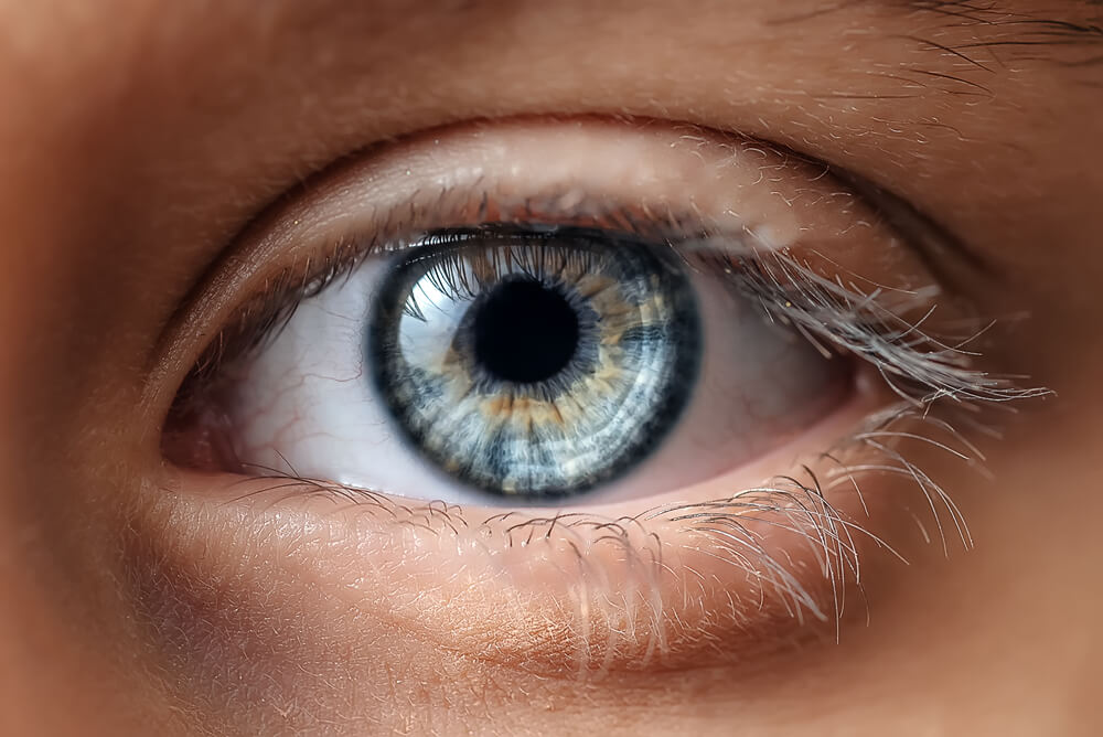
Retinal conditions we treat
Our retina specialists are experts in diagnosing and treating various retinal conditions. Some of the commonly encountered retinal conditions include:
- Age-related macular degeneration: This condition primarily affects older adults and can cause central vision loss, impairing activities like reading and recognising faces.
- Retinal detachment: A serious condition in which the retina separates from the back of the eye, often caused by trauma or other underlying eye disorders. Immediate medical attention is necessary to prevent permanent vision loss.
- Diabetic-related eye conditions: Individuals with diabetes are at risk of developing retinopathy, a condition that damages the blood vessels in the retina. Diabetic retinopathy can lead to vision impairment if left untreated.
- Uveitis: This inflammatory condition affects the eye’s middle layer, including the retina. Prompt treatment with a Uveitis specialist is crucial to prevent potential vision loss and complications.
- Macular hole: A macular hole is a small break in the macula, the central part of the retina responsible for sharp, detailed vision. Surgical intervention may be necessary to restore vision in severe cases.
- Floaters: Floaters are specks or cobweb-like shapes that appear to drift across your field of vision. While they are usually harmless, sudden changes in the number or size of floaters may indicate a retinal issue that requires attention.
If you experience symptoms such as sudden vision changes, flashes of light, or new floaters, consult a retina specialist. Early detection and appropriate treatment can significantly improve outcomes for retinal conditions.
At Moorfields Eye Hospitals, our Retina and Uveitis specialists in Dubai and Abu Dhabi employ state-of-the-art diagnostic tools and advanced treatment techniques to preserve and restore your precious eyesight.
Looking for more information?
Explore our frequently asked questions for additional information about your condition, care and treatment.
Conditions & Treatment

Macular Hole Surgery
About Macular Hole Surgery
If you think of your eye as a camera, the retina is like the photographic film. It is a very thin layer of tissue, which is sensitive to the image focused on it, sending the information to the brain.
At the very centre of the retina is the macula. This is a very special area of the retina, which we use for reading and recognising complex shapes. Sometimes, a hole forms in the macula, which prevents it from working normally. This affects your vision, particularly for reading and other visually demanding tasks, but it does not cause total blindness.


Posterior Vitreous Detachment
About Posterior Vitreous Detachment
Posterior Vitreous Detachment (PVD) is a common degenerative change which affects one or both eyes in many people after middle age. It may present earlier in short-sighted patients or those who have sustained trauma to the eyes.
Thickening of the jelly casts shadows on the retina and is seen as floating shapes. These black 'floaters' in your vision move with the eye and then settle as the eye rests. These are often described by patients as a “cobweb” or “insects”.
You may also notice flashing lights, like little flickers in the outer periphery. Usually, these do not indicate a problem; however, it is important to have the eye thoroughly checked, as a retinal tear or retinal detachment may occasionally occur.

Retinal Detachment Surgery
About Retinal Detachment Surgery
Retinal detachment is a condition in which the thin lining at the back of the eye (the retina) begins to come away and separate itself from the underlying wall of the eye, which contains blood vessels that supply it with vital oxygen and nutrients.
If not treated promptly, retinal detachment will lead to blindness in the affected eye.
A retinal detachment is usually caused by a tear in the retina, and this is termed a Rhegmatogenous retinal detachment. Other types include traction retinal detachment, usually seen in advanced diabetic retinopathy, and Exudative retinal detachment (usually seen in people with inflammation). Rhegmatogenous retinal detachment needs urgent surgery in most cases.

Retinal Photodynamic Therapy (PDT)
About Retinal Photodynamic Therapy (PDT)
Photodynamic therapy (PDT) is a treatment that uses a combination of a “cold” laser and a special light-sensitive dye (Verteporfin). This is injected into the bloodstream to target abnormally leaking blood vessels in the retina (nerve tissue lining at the back of the eye which detects light and allows us to see) or the layer below the retina (choroid). PDT is used to treat some specific forms of wet macular degeneration and a disease called ‘Central Serous Retinopathy’ (CSR).

Retinal Vein Occlusion
About Retinal Vein Occlusion
Blocking of the retinal vein, which reduces vision, is known as Retinal Vein Occlusion. This results in the accumulation of blood (retinal haemorrhages) and fluid (macular oedema) in the retina and leads to a drop in visual acuity (clarity of vision).

Diabetic Retinopathy
About Diabetic Retinopathy
Diabetic retinopathy is a serious complication of long-term uncontrolled diabetes resulting from persistently high blood sugar levels over the years. This condition affects the retina, the light-sensitive tissue at the back of the eye, impairing its ability to transmit clear images to the brain through the optic nerve.
At Moorfields Eye Hospitals, our retina specialists in Dubai and Abu Dhabi have extensive subspecialty training in diagnosing and treating diabetic retinopathy. Using the latest retinal imaging and advanced treatment approaches, they provide tailored care to slow disease progression, protect sight, and help patients maintain long-term eye health and quality of life.

Diabetic Maculopathy
About
Diabetic maculopathy is a condition that occurs when the macula, the part of the eye which provides us with our central vision is damaged. The macula is responsible for fine vision (reading, writing, watching television, and recognising faces). Patients with diabetes may develop macular oedema (swelling of the retina) due to leaking of fluid from blood vessels. This causes the vision to become blurred.

Age-related Macular Degeneration (AMD)
About Age-related Macular Degeneration (AMD)
Age-related macular degeneration (AMD) is an eye condition that affects the macula, a small area at the centre of the retina responsible for sharp, detailed vision. AMD becomes more common with age and can affect one or both eyes.
AMD can cause blurred, distorted or reduced central vision, making everyday activities such as reading, driving and recognising faces more difficult. Straight lines may appear wavy, and in advanced cases, dark or blurred areas may develop in the centre of vision. Peripheral (side) vision usually remains unaffected.
In its early stages, AMD may develop without noticeable symptoms, making regular eye examinations important for early detection and monitoring.
Retina services Specialists

Dr. Ammar Safar
Chief Medical Officer and Consultant Ophthalmologist & Vitreoretinal SurgeonGCAA Approved Specialist Aviation Medical Examiner
Professor of Ophthalmology (Adjunct)
- Vitreoretinal diseases and surgery
- Retinal Detachment surgery
- Diabetic retinopathy
- Macular hole surgery
- Cataract surgery
- Intravitreal injections
- Retinal vascular diseases and vascular occlusive disorders
- Age-related macular degeneration, including exudative macular degeneration
- Aviation Ophthalmology
- American Board-Certified Ophthalmologist
- Medical degree from Aleppo University, Aleppo, Syria
- Residency training in Ophthalmology at Georgetown University, Washington, DC, USA
- Served as Chief Resident at Georgetown University, Washington, DC, USA
- Fellowship in Vitreoretinal Diseases & Surgery at the University of Texas Southwestern Medical Centre, Dallas, Texas, USA
- American Academy of Ophthalmology
- Fellow of the American College of Surgeons (FACS)
- American Society of Retina Specialists
- European Society of Retina Specialists
- American Society of Cataract and Refractive Surgery
- Association for Research in Vision and Ophthalmology (ARVO)
- Arab-American Medical Association
- Adjunct Professor of Ophthalmology at Mohammed Bin Rashid University of Medicine and Health Sciences (MBRU)
- Former Assistant Professor and later Associate Professor and Director of Vitreoretinal Surgery at the Department of Ophthalmology, Jones Eye Institute, University of Arkansas for Medical Sciences, USA
- Conducted research and clinical trials in diabetic retinopathy and age-related eye diseases
- Research contributed to advances in the management of retinal vascular diseases, including diabetic retinopathy and vascular occlusive disorders
- Research contributed to new approaches for the treatment of exudative macular degeneration
- Inaugural recipient of The Martha Wood Bentley Endowed Chair of Ophthalmology at the University of Arkansas


Dr. Fahd Quhill
Consultant Ophthalmologist in Medical Retina and Ocular inflammatory disease (Uveitis)Clinical Professor Of Ophthalmology (Adjunct)
- Uveitis and ocular inflammatory diseases
- Infectious eye diseases
- Autoimmune diseases affecting the eye
- Complex inflammatory eye disease requiring immunosuppressive treatment
- Diabetic retinal disease
- Age-related macular degeneration
- Inherited retinal diseases
- Retinal vascular diseases
- Bachelor of Medicine and Bachelor of Surgery (MBChB), University of Birmingham, UK
- Fellow of the Royal College of Surgeons of Edinburgh (FRCSEd)
- Member of the Royal College of Ophthalmologists (MRCOphth)
- Bachelor of Medical Science (BMedSc), University of Birmingham, UK
- Ophthalmology specialist training in Oxford, Birmingham and London, UK
- Three Medical Retina Fellowships completed in Birmingham, Wolverhampton and Moorfields Eye Hospital London, UK
- Member of the Royal College of Ophthalmologists
- Clinical Professor of Ophthalmology (Adjunct)
- Principal Investigator in numerous clinical trials investigating macular degeneration, retinal vein occlusion and diabetic macular oedema
- Author of 27 research papers on retinal and ocular inflammatory diseases
- Research published in leading peer-reviewed journals, including Ophthalmology, British Journal of Ophthalmology and Eye

Dr. Avinash Gurbaxani
Visiting Consultant Ophthalmologist in Uveitis and Medical Retinal Diseases and Cataract Surgery- Uveitis and inflammatory eye diseases
- Complex inflammatory eye diseases requiring immunosuppressive treatment
- Diabetic retinal disease
- Age-related macular degeneration
- Retinal vascular diseases
- Medical retinal diseases
- Cataract surgery including premium intraocular lenses
- Complex cataract surgery associated with inflammatory and retinal eye diseases
- Bachelor of Medicine and Bachelor of Surgery, India
- Diploma in Ophthalmic Medicine and Surgery, India
- Fellowship of the Royal College of Surgeons of Edinburgh in Ophthalmology
- Ophthalmology training in Oxford and London, UK
- Medical Retina and Uveitis Fellowship at Moorfields Eye Hospital, London, UK
- Medical Retina and Uveitis Fellowship at Sydney Eye Hospital, Australia
- Royal College of Ophthalmologists (RCOphth)
- Royal College of Surgeons of Edinburgh (RCS Ed)
- American Academy of Ophthalmology (AAO)
- Experienced in initiating ophthalmology research projects
- Experience in conducting clinical trials
- Regularly publishes research papers.

Dr. Muhammad Amjad
Visiting Consultant Ophthalmologist in Adult and Paediatric Retinal diseases and Cataract surgery- Adult and paediatric retinal diseases
- Retinal detachments
- Macular holes
- Age Related Macular Degeneration
- Retinal vascular diseases
- Epiretinal Membranes
- Vitreous Haemorrhage
- Uveitis
- Inherited retinal disorders (IRD)
- Retinopathy of Prematurity (ROP)
- Central Serous Chorioretinopathy (CSCR)
- Familial Exudative Vitreoretinopathy (FEVR)
- Adult and paediatric cataract surgery
- Bachelor of Medicine and Bachelor of Surgery (MBBS), University of Health Sciences, Lahore, Pakistan
- Certificate of Completion of Training (CCT), General Medical Council, UK
- Fellowship in Medical Retina, Uveitis & Oncology at Moorfields Eye Hospital London, UK
- Fellowship in Surgical Retina at Moorfields Eye Hospital London, UK
- Fellowship in Paediatric Retina at Great Ormond Street Hospital for Children, London, UK
- Fellow of the Royal College of Ophthalmologists (FRCOphth)
- British and Eire Association of Vitreoretinal Surgeons (BEAVRS)
- European Society of Retina Specialists (EURETINA)
- American Society of Retina Specialists (ASRS)
- American Academy of Ophthalmology (AAO)
- British & Irish Paediatric Ophthalmology & Strabismus Association (BIPOSA)
- Active involvement in retinal research
- Published peer-reviewed research on Retinopathy of Prematurity (ROP) and Familial Exudative Vitreoretinopathy (FEVR)
- Research published in international ophthalmology journals
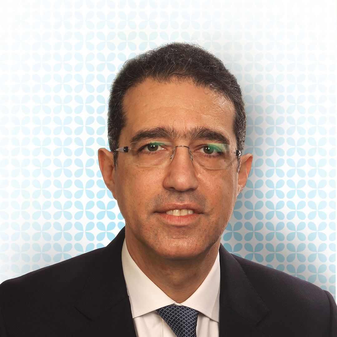
Dr. Ziad Bashshur
Visiting Consultant Ophthalmologist in Medical & Surgical Retina- Medical retina
- Surgical retina
- Vitreoretinal diseases and surgery
- Doctor of Medicine (MD) from the American University of Beirut, Lebanon
- Internal Medicine internship at Georgetown/VA Medical Center, USA
- Ophthalmology residency at the American University of Beirut, Lebanon
- Vitreoretinal Fellowship at the University of Virginia, USA
- Member of the American Society of Retina Specialists (ASRS)
- Member of the Retina Society
- Member of the Alpha Omega Alpha Honor Medical Society
- Tenured Professor and Vice-Chairman of Ophthalmology at the American University of Beirut
- Published more than 60 research papers on retinal diseases
- Research areas include macular degeneration, diabetic retinopathy, retinal vascular diseases and retinal imaging
- Regular speaker at professional ophthalmology meetings worldwide
- Serves on the review boards of several peer-reviewed ophthalmology journals
- Serves on several medical industry advisory boards

Dr. Wajiha Kheir
Visiting Consultant Ophthalmologist in Medical Retina and Ocular Oncology- Medical retina
- Ocular oncology
- Age-related macular degeneration
- Radiation retinopathy
- Retinal degenerations
- Electrophysiology of vision
- Bachelor of Science in Biology with High Distinction from the American University of Beirut, Lebanon
- Recipient of the Penrose Award
- Doctor of Medicine (MD) from the American University of Beirut, Lebanon
- Internal Medicine internship at the American University of Beirut Medical Center, Lebanon
- Ophthalmology residency at the American University of Beirut Medical Center, Lebanon
- Fellowship in Medical Retina at Duke Eye Center, Duke University School of Medicine, USA
- Fellowship in Ocular Oncology with Clinical Pathology Concentration at Duke Eye Center, Duke University School of Medicine, USA
- Alpha Omega Alpha (AOA) Honor Medical Society
- American Academy of Ophthalmology (AAO)
- International Society of Ocular Oncology (ISOO)
- Assistant Professor of Ophthalmology at American University of Beirut
- Author of 18 peer-reviewed journal articles
- Author of 10 book chapters
- Research presented in more than 40 poster and paper presentations at regional and international conferences
- Research interests include age-related macular degeneration, Ocular oncology, radiation retinopathy, retinal degenerations and electrophysiology of vision

Dr. Usman Mahmood
Consultant, Ophthalmologist Specialised in Retina and Cataract Surgery
FAQ
Retinal conditions can cause blurred or distorted vision, difficulty seeing fine detail, flashes of light, new or increasing floaters, reduced peripheral vision or a shadow across part of the visual field. Some retinal conditions, including early diabetic retinopathy, may develop without noticeable symptoms, which is why regular assessment with a retina specialist is important for patients with known risk factors.
Floaters are common and are often harmless, but a sudden appearance or increase in floaters should be assessed urgently, particularly when accompanied by flashes of light, blurred vision, a shadow or curtain across the vision, or sudden loss of sight. These symptoms can indicate a retinal tear or retinal detachment, which requires prompt examination and treatment.
Diabetes can lead to diabetic retinopathy, which can develop without noticeable symptoms during its early stages, so changes to vision are not a reliable indication that the retina is unaffected. Patients with diabetes should undergo retinal assessment at the intervals recommended by their treating clinician, allowing changes to be identified and managed before they become sight-threatening.
A retinal assessment usually includes checking your vision and examining the back of the eye after the pupils have been dilated. Depending on the findings, further investigations may include retinal photography, optical coherence tomography (OCT), OCT angiography, dye-based angiography, among other tests, with each test selected according to the condition being investigated.
Treatment depends on the condition, its severity, and its effects on the retina and vision, and may include regular monitoring, injections into the eye, laser treatment, photodynamic therapy, or retina surgery. Our retina specialist in Dubai and Abu Dhabi will explain the clinical findings, the purpose of the recommended treatment and what outcome can reasonably be expected before care is planned.
We have medical and surgical retina specialists in Dubai and Abu Dhabi who diagnose and manage a wide range of retinal conditions, both routine and complex. As we provide care across all major ophthalmic specialities, patients can also be referred directly to another subspecialist when their condition affects more than one part of the eye or requires a coordinated approach.
Our retina specialists in Dubai and Abu Dhabi diagnose and treat a wide range of conditions affecting the retina, including age-related macular degeneration, diabetic retinopathy, retinal detachment, retinal tears, macular holes, retinal vein occlusion, and floaters.
Retinal care at our specialist eye hospitals in Dubai and Abu Dhabi is delivered within established clinical governance, audit and patient safety frameworks, with protocols aligned to those followed by Moorfields Eye Hospital London and supported by JCI and ACHSI accreditation. We regularly measure and publish clinical outcomes to provide patients with clear, transparent information about the quality of care we deliver.
Learn more about our satisfaction rates and outcomes: https://moorfields.ae/our-satisfaction-rates/