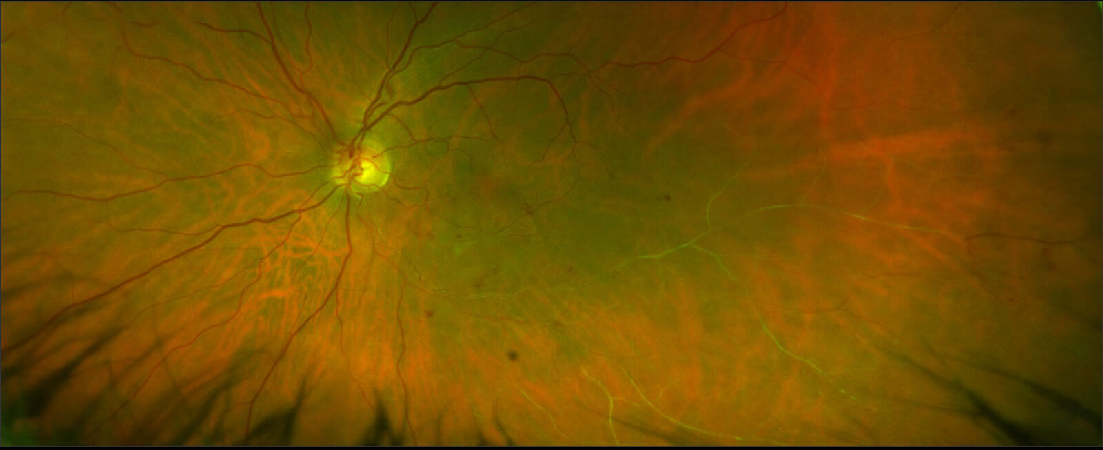
About Retinal Vein Occlusion
Blocking of the retinal vein which reduces the vision is known as Retinal Vein Occlusion. This results in the accumulation of blood (retinal hemorrhages) and fluid (macular edema) in the retina and leads to a drop in the visual acuity (clarity of vision).
Types of Retinal Vein Occlusion
There are two types of Retinal Vein Occlusion:
- Central Retinal Vein Occlusion (CRVO)
- Branch Retinal Vein Occlusion (BRVO)
Causes
A blockage forms in the vein, usually due to a blood clot, and obstructs the blood flow. The exact cause is unknown, but several conditions make the condition more likely. These include:
- High blood pressure
- High cholesterol
- Glaucoma
- Diabetes
- Smoking
- Certain rare blood disorders
Symptoms
Retinal vein occlusion sometimes may not have any symptoms. However, some symptoms to observe are:
- Blurry or missing vision in part or all of an eye
- Dark spots or lines floating the vision
- Pain and pressure in the eye
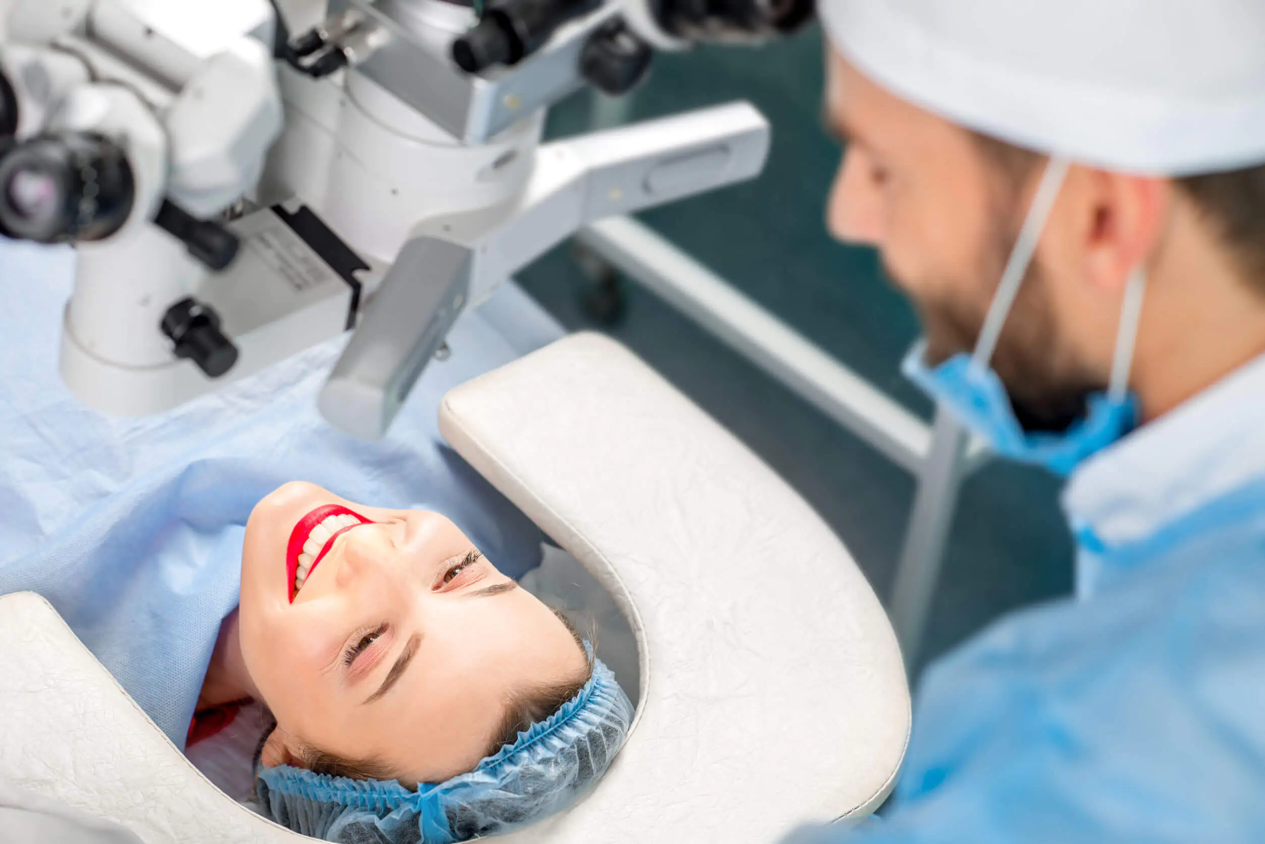
Diagnosis
Retinal Vein Occlusion is diagnosed clinically with a dilated eye examination. Additional imaging such as fluorescein angiography and ocular coherence tomography (OCT) may be needed to confirm the diagnosis and monitor the progression of the disease.
Treatment of Retinal Vein Occlusion includes:
Ocular treatment:
- Observation if only a small vein is involved and does not affect the macula (central part of the vision).
- Intravitreal injections of either anti-vascular endothelial growth factor (anti-VEGF) agents or steroid injection to treat the macular edema.
- Laser photocoagulation may be administered to the peripheral ischemic retina.
Systemic treatment:
- Management of systemic factors (close control of blood pressure and blood sugar levels).
Retinal Vein Occlusion Specialists

Dr. Ammar Safar
Chief Medical Officer and Consultant Ophthalmologist & Vitreoretinal SurgeonGCAA Approved Specialist Aviation Medical Examiner
Professor of Ophthalmology (Adjunct)

Dr. Fahd Quhill
Consultant Ophthalmologist in Medical Retina and Ocular inflammatory disease (Uveitis)Clinical Professor Of Ophthalmology (Adjunct)

Dr. Luisa M. Sastre
Consultant Ophthalmologist in General Ophthalmology and Cataract SurgeryGCAA Approved Specialist Aeromedical Examiner
Dr. Avinash Gurbaxani
Visiting Consultant Ophthalmologist in Uveitis and Medical Retinal Diseases and Cataract Surgery
Prof. Ziad Bashshur
Visiting Consultant Ophthalmologist in Medical & Surgical Retina
Dr. Wajiha Kheir
Visiting Consultant Ophthalmologist in Medical Retina and Ocular Oncology