Diabetic Retinopathy

This week’s blog on Diabetic Retinopathy has been contributed by Dr Paola Salvetti, Consultant Ophthalmologist and Specialist in Medical Retina.

Diabetes

Diabetes is a disease related to the body’s inability to properly absorb, store and use sugar from food. This results in higher levels of sugar in the blood (hyperglycaemia).

Differences between type 1 and type 2 diabetes

Type 1 diabetes is often referred to as ‘insulin dependent’ and is mostly diagnosed in children or young adults. Type 2 diabetes is the most common type; it usually appears in adults over the age of 40 and is often related to overweight, obesity and lack of physical activity in people who are genetically predisposed.
Genetic predisposition affects people differently, depending on the type:

  • In type 2 diabetes, if one parent is affected the risk of transmission to children is about 40%, which increases to 70% if both parents have the disease.
  • In type 1 diabetes, the risk is much lower, being only 5%.

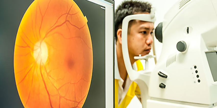
Diabetes is a significant public health issue in the region, with the UAE, Saudi Arabia, Bahrain, Kuwait all featuring in the top 15 countries in terms of the prevalence of diabetes worldwide. According to data from The Institute for Health Metrics and Evaluation from 2015, 19.3% of the UAE population between the ages of 20 and 79 have type 2 diabetes, which is almost 1 in 5 people, meaning that there are over 1 million people living with diabetes in the UAE. In the UAE, diabetes is also among the top seven causes of premature death and is 4th among the top causes of disability; diabetes increased by a staggering 174% between 2005 -2015.
Type 2 diabetes can sometimes be difficult to diagnose, as it is often painless and without symptoms, at least at the beginning. In fact, it is estimated that the interval between the first appearance of hyperglycaemia and the diagnosis of diabetes is around 5 to 10 years.
Prolonged and repeated hyperglycaemia leads ultimately to the damage of blood vessels and nerves throughout the body, and this includes complications in the eyes, kidneys, heart, brain and limbs.

Diabetic Retinopathy

Diabetic Retinopathy is one of the most common and serious complications of diabetes and its prevalence increases with the amount of time diabetes is present, with age, poor control of blood sugar, blood pressure, cholesterol and lipids.
Although some visual problems can indicate the presence of Diabetic Retinopathy (such as blurred letters when reading, vision difficulties when moving from light to dark) more commonly the disease starts in a completely silent way with no obvious symptoms.
Diabetic Retinopathy can be present even in patients with excellent visual acuity and no symptoms, and can only be diagnosed by the ophthalmologist with a Fundus examination. Sometimes, additional tests are required, such as OCT and Fluorescein Angiography to evaluate the risk of the progression of retinopathy. Some early cases can be just observed, whilst more advanced cases require active management.

Diabetic Retinopathy Treatment

Different treatment options are available, and sometimes a combination of the possible treatments is used. These include intravitreal injection of medications (anti VEGF and or steroids), conventional peripheral laser, subthreshold micropulse laser. In more advanced cases, with prolipherative retinopathy or with intravitreal haemorrhages, surgery may be the only possible option.
Early diagnosis of the complications of diabetes generally leads to better management and better outcomes, and that is why it is vitally important to make regular visits to the ophthalmologist.

Keratoconus

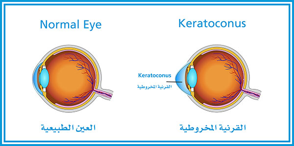
This blog on Keratoconus has been contributed by Dr Osama Giledi, Consultant Ophthalmologist, Specialist in Cataract, Cornea and Refractive Vision Correction Surgery

What is Keratoconus?

The word Keratoconus comes from two Greek words: kerato (cornea) and konos (cone). Keratoconus is a degenerative disorder of the eye in which the shape of the cornea, which is usually round, is distorted and develops a cone-shaped bulge, resulting in reduced vision. Keratoconus is a relatively common condition. It affects one person in two thousand, occurs around the world and is quite common in the Middle East.
Progression of Keratoconus depends on the patient’s age at the time of onset and the severity of eye rubbing. The earlier the onset, the faster Keratoconus may progress. The condition always affects both eyes and is asymmetric, so one eye may be more affected than the other.

Causes

Keratoconus has many causes, and it results in a cornea that is more elastic than normal and that starts to thin and bulge forward, causing reduced vision and increased astigmatism. It can be inherited, and a chromosomal link has been identified. Keratoconus can be associated with other allergic diseases such as hay fever, eczema and asthma but we are not born with it, and its onset usually happens around puberty. It is strongly associated with eye rubbing, making it progress rapidly. It is also seen in contact lens wearers.
For the best outcome, Keratconus should be detected and treated early. Early detection is possible by generating computerized corneal shape and thickness measurements through corneal topography using advanced technology such as a pentacam machine. If left untreated, Keratoconus could lead to significant visual impairment and blindness.

Treatment

Spectacles
These can provide good vision in the early stages of the condition and usually help to correct myopic astigmatism.
Hard or Scleral Contact Lenses
When spectacles don’t work, rigid contact lenses and Scleral lenses usually provide very good vision if the patient can tolerate them; they should be fitted by a contact lens specialist.
CXL – Corneal Collagen Cross linking with Riboflavin (Vitamin B2)
This technique increases the strength of the cornea and prevents the progress of Keratoconus.
Intracorneal Rings (Intacs & Ferrara)
These rings inserted into the cornea change the shape of the corneal cone, flattening it and making it a more regular and central shape. This will help improve the vision for the majority of patients.
We use it when patients cannot tolerate contact lenses and while the cornea is still clear and the condition is not very advanced. A Femto-second laser is used to create the tunnel and the entry point for the rings, making it a very safe and reliable procedure.
Toric Implantable Contact Lenses
For those with stable Keratoconus and good vision with glasses, Toric Implantable Contact Lenses are generally used (sometimes after stabilizing the Keratoconus with corneal cross linkage) to improve corneal shape with an intracorneal ring or with very limited therapeutic Excimer laser correction. The Toric ICL can eliminate the need for optical aids altogether and, in some patients, results in improved vision and balance between the two eyes.
Phototherapeutic Keratectomy (PTK)
Keratoconus patients are not candidates for Lasik or normal laser correction. However, some patients benefit from limited excimer laser correction to improve the surface irregularity of the cornea when
contact lenses cannot be tolerated. PTK is always combined with corneal cross-linkage, undertaken during the same procedure or afterwards.
Corneal Transplants
Corneal Transplants are used in the advanced stages of Keratoconus, when there is corneal scarring. The procedure is either a partial thickness (Deep Anterior Lamellar Keratoplasty = DALK) or full thickness graft (Penetrating Keratoplasty = PK). The need for corneal grafts is declining because of the increasing success in the early diagnosis of Keratoconus and the effectiveness of corneal cross linkage to stabilize the condition, so Keratoconus generally does not reach the advanced stage.

Refractive Surgery

This week’s blog on refractive surgery has been contributed by Dr Hamed Mofeez Anwar, Consultant Corneal and Refractive Surgeon.

If you’re nearsighted, farsighted, have astigmatism or are presbyopic (loss of reading ability due to age), refractive surgery is a term referring to procedures which can be used to improve your vision.

What are the benefits of refractive surgery?

For most patients, the vision after refractive surgery is similar to that with contact lenses prior to surgery, without the potential discomfort and limitations of performing activities.

Over 95% of patients are satisfied with the outcome of the surgery, with many describing it as a “life-changing”. Although refractive surgery is often considered to be a cosmetic surgery procedure, the benefits are primarily functional. It’s designed to make you less dependent on glasses and contact lenses, letting you lead an active lifestyle more easily.

Am I a good candidate for refractive surgery?

Refractive surgery may be a good option for you if you:

  • Want to decrease your dependence on glasses or contact lenses.
  • Are free of eye disease.
  • Accept the inherent risks and potential side effects of the procedure.
  • Understand that you might still need glasses or contacts after the procedure to achieve your best vision.
  • Have an appropriate refractive error

What types of refractive surgery are there?

It’s important to remember that there is no universally-accepted, best method for correcting refractive errors.

The best option for you should be decided after a thorough examination and discussion with your ophthalmologist, especially taking into account your lifestyle and vision needs.

The focusing ability of your eyes can be adjusted by procedures which include:

  • Laser vision correction – these procedures adjust the focusing power of the eye by reshaping the cornea (the clear window at the front of the eye).
  • Implanting a lens inside the eye.

Laser refractive surgery:

Laser vision correction procedures use an excimer laser to reshape the cornea (the clear window at the front of the eye), therefore correcting refractive errors. Generally speaking, the cornea is flattened to treat near-sightedness or steepened to correct far-sightedness.

Laser refractive surgery procedures include:

  • LASIK
  • Surface laser treatment (PRK/Trans-PRK/LASEK)

The risks and benefits are similar amongst the two procedures and they generally provide good results in the appropriate patients.

The main difference among the two procedures is the speed of recovery. LASIK patients are usually able to return to work within a day or so after surgery while patients who have PRK done may need up to a week to attain driving standard vision.

It’s important to keep in mind that the visual results at 3 months are equivalent for all types of surgery.

LASIK

In LASIK, a very thin flap is created on the surface of the cornea using a femtosecond laser. This flap is then lifted up and an excimer laser is then applied to the corneal surface. At the end of the procedure, the flap is then placed back into its original position. After surgery, a minimal amount of discomfort may be experienced. Visual recovery after surgery is rapid, with many patients seeing well enough to work and drive within a day or so. However, it’s important to remember that patients who engage in contact sports must wait a month before resuming activities.

This video http://fyi.rendia.com/XZN8k shows how LASIK eye surgery is performed.

Surface laser treatment (PRK/TransPRK/LASEK)

In PRK laser treatment, a laser is applied directly to the corneal surface. This removes a lens shaped piece of tissue immediately below the clear skin of the cornea. The skin regrows over the course of a week, and then smooths out over the next 3 months. While the skin is growing again, the eye surface is normally very sore. This is the main difference between surface laser treatments and LASIK, both of which aim to keep the corneal skin layer intact.

This procedure requires minimal surgical manipulation and is especially suited to patients that aren’t suitable for LASIK because of a thin cornea or a very active lifestyle that puts them at risk of dislodging a corneal flap. The downsides of this procedure are postoperative pain that lasts a day or so after surgery and a more prolonged visual recovery that would extend to a week to attain driving standard vision. Patients who engage in contact sports can resume activity much sooner than with LASIK.

Laser vision correction for decreased reading vision (presbyopia)

Age related loss of reading vision (presbyopia) can be treated with laser vision correction surgery. To do this, one eye is treated so it sees well for distance, while the other eye is treated to see well for closer objects. This type of treatment is called monovision. When both eyes are open, our brain combines the two images to partially restore near vision with little compromise optically. This is the default strategy for improving the near range in older patients undergoing laser vision correction.

Lens implantation surgery:

Laser vision correction is not suitable for all patients.

Some people have high degrees of refractive errors which can’t be safely corrected with laser-based refractive surgery. Other people (generally above the age of 50) may have early cataractous changes in the lens of their eyes.

Younger patients:

In younger patients, a lens is surgically implanted inside the eye in front of the natural lens. This lens is placed either in front of or behind the iris of the eye. Once the lens is properly positioned inside the eye, it provides the necessary correction to focus light rays onto the retina. It’s important to note that the natural lens of the eye is not removed during this procedure.

Older patients

Early stages of cataract are often seen in older patients, and lens replacement surgery may be more appropriate in this group. In this surgery, the natural lens is replaced with a lens implant. A variety of different implants are used, which include multifocal lenses designed to reduce reliance on spectacles for near, intermediate and distance vision.

Is refractive surgery permanent?

In most cases, yes. Short sight and astigmatism normally stabilise in the late teens or early 20s, however natural changes in your eye power can happen at any stage in life. As a result, laser vision correction sometimes needs to be repeated.

Uveitis

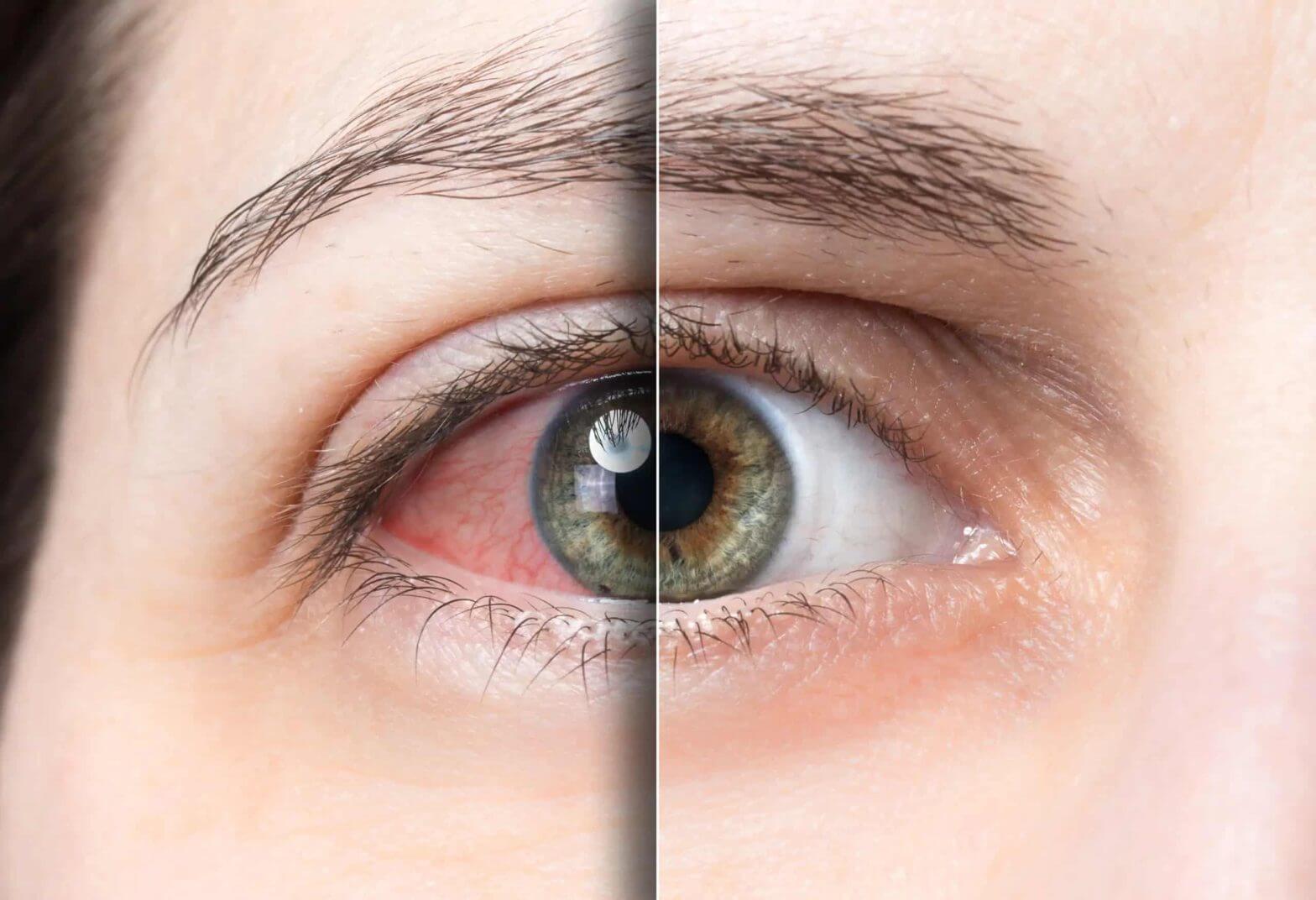
This week’s blog on Uveitis has been contributed by Dr Avinash Gurbaxani, Consultant Ophthalmic Surgeon in Uveitis and Medical Retinal Diseases and Cataract Surgery.
Uveitis is not a single disease but a clinical spectrum of symptoms and signs caused by a variety of medical conditions. Although it accounts for about 1% of all eye diseases, Uveitis is the cause of 10-15% of blindness, thus, it must be managed very carefully. Prompt and appropriate treatment is needed to ensure good visual recovery, which is very often achievable.  In most cases, a Uveitis specialist – an eye doctor with specialist training in diagnosing and managing these diseases – should manage Uveitis.
Uveitis can affect children, especially those with childhood arthritis or infections. Uveitis affects young adults of a working age and more often women are affected. Symptoms include red eye (although in children the eyes do not become red, so if diagnosed with arthritis, they must have regular screening with the Uveitis specialist), pain, light sensitivity, and blurred vision. It may be associated with skin rashes, joint pains, and stomach symptoms.

Causes:

The cause of Uveitis can be infection (like TB, Toxoplasmosis or viruses like herpes and CMV) or autoimmune conditions (sarcoidosis, arthritis, inflammatory bowel disease). In about half of the cases, we may never find a specific cause but prompt treatment is always needed.
Initially, patients will often need to have a variety of blood tests to look for infection or auto immune conditions, chest x-rays and other imaging. In the eye clinic patients may need an OCT scan of the retina and a flurosein angiogram to look for leakage from the blood vessels in the retina. Patients may also be referred to othzer specialists like rheumatologists, gastroenterologists or neurologist to help diagnose a systemic cause of the Uveitis.

Types and treatment:

Uveitis can be in the anterior segment (front part of the eye), which usually can be treated with drops and sometimes injections to get a quick response. When Uveitis affects the back of the eye, more aggressive treatment may be needed. This usually consists of a high dose of oral steroid medications for several weeks. Antibiotics are given in case of infection. Long-term treatment or more serious cases may need steroid sparing immune suppressant medication. This scan be in the form of tablets like mycophenolate or methotrexate or newer biologic medications, which are given in the form of injections. Steroid injections into the eye may be needed as well. Although protracted, early, aggressive and appropriate treatment can achieve good control of this condition and patients can maintain good vision.

Complications:

Complications of Uveitis include cataract, high pressure in the eye and macular oedema. These may need additional treatment including surgery.
If you have been diagnosed with Uveitis or suspect that you may have Uveitis, it is essential to see a Uveitis specialist