


About Visual Electrophysiology
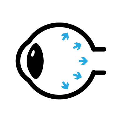
When we look at something, a picture of the object is projected onto the retina at the back of our eye. The retina changes this optical picture into little electrical signals, which pass along the optic nerve to the brain, where the sense of ‘seeing’ happens. Visual electrophysiology measures these small signals created by the eye and the brain.
Visual electrophysiology assesses how the visual system processes visual information. The tests support the diagnosis of the visual problem. They are also useful for monitoring the progression of a visual disorder or the effects of any treatment. These tests are particularly useful in young infants and children as they sometimes are unable to communicate or describe in detail any problems they might have with how they see.
How are the tests conducted?
Small electrical signals from the eye are recorded in response to a flashing light or a pattern on a computer screen. These signals are recorded by small contacts that are placed on the surface of the head and near the eye. The area under the eye will be gently cleaned with a gel before the contact is applied. This may be slightly uncomfortable but not painful. For some of the tests, we may need to use dilating eye drops, similar to those used in the clinic, or very occasionally, anaesthetic (numbing) drops.
How should I prepare myself for the tests?
The tests are noninvasive but require small contacts to be placed around or in the eyes as well as around the head. Before attending your appointment, please ensure your face and skin are free of creams, oils and makeup.
Services offered
Visual evoked potential (VEP):
Recording the brain’s activity is called the visual evoked potential (VEP). To record the VEP, small contacts are placed on the patient’s head, using some paste. You will be asked to look at a moving black-and-white pattern on a TV screen and a light which will flash twice a second.
Electroretinogram (ERG):
The recording of the retina’s activity is performed using the electroretinogram (ERG). To record the ERG, dilating eye drops are used. In adults and older children, small contacts are placed at the side of the eye and over the lower eyelid. In babies and young children, the contacts are placed at the side of the eye and below each eyelid and are held by some tape. You will be asked to look at a light that flashes at different speeds and brightness levels.
Pattern Electroretinogram (PERG)
The pattern electroretinogram (PERG) records the activity of the central macula. To record the PERG, small contacts are placed at the side of the eye and over the lower eyelid. Anaesthetic (numbing) drops will be instilled before applying the contacts over the lower eyelid. You will be asked to look at a moving black-and-white pattern on a screen.
Multi-focal Electroretinogram (mfERG)
The multifocal electroretinogram (mfERG) test examines specific areas or focal points within the retina. Small contacts are placed at the side of the eye and over the lower eyelid, similar to the ERG and PERG. Anaesthetic (numbing) drops will be used before applying the contacts over the lower eyelid. You will be asked to look at a moving hexagonal pattern on a screen.
Electro-oculogram (EOG)
The electrooculogram (EOG) test specifically measures the function of the retinal pigment epithelium layer (RPE). This very important layer nourishes and supports the normal functioning of the retina. Small contacts are placed around the side of the eyes, and you will be asked to follow a moving light from side to side. Dilating drops will be used, and the test is performed in the dark and the light.
