Diabetic Maculopathy

Intravitreal Injection Treatment

The macula is the central part of the retina at the back of the eye. It is responsible for fine vision (reading, writing, watching television, and recognising faces). Patients with diabetes may develop macular oedema (swelling of the retina) due to leaking of fluid from blood vessels. This causes the vision to become blurred.
For some patients a course of anti VEGF injections into the eye will be needed to treat the swelling and improve the vison. The drug stops the growth of the abnormal blood vessels and reduces the swelling of the retina.
A course of three injections is recommended each injection one month apart at the start. The procedure is carried out in a clean environment using sterile technique.
The eye is cleaned and local anaesthetic drops are given to numb the eye. During the injection you will feel a sensation of pressure but it should not be painful. It takes less than a minute to give the injection and the needle is slowly withdrawn when finished.
Afterwards the eye may feel slightly bruised and a bruise may appear on the white of the eye. This is not a serious problem and is due to blood vessels in the lining of the outside of the eye being caught by the needle.
This will slowly disappear over a couple of weeks. If your eye becomes very red and/or painful in the days and weeks following the injection you should contact the hospital.
The risks of the injection are bleeding and infection. The risk of this happening is one person in every 3,000.

Aftercare

The eye may or may not be covered after the injection. If a pad is applied this may be removed when you reach home.
We do not usually give post injection drops. If the eye is uncomfortable you may take 2 paracetamol or your regular pain relief tablet.

  • Do not splash water into the eye for one day.
  • You may perform all your usual activities from the following day.
  • When ready for discharge you will be given an appointment for a follow up visit.

Meibomian Gland Disfunction (MGD)

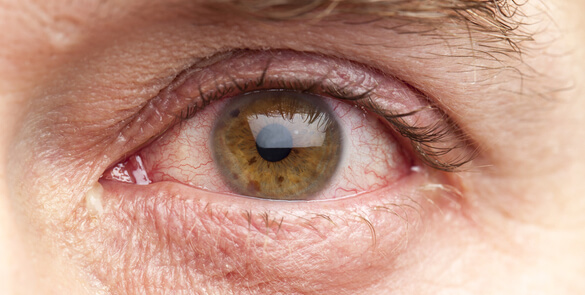
What is Meibomian Gland Disfunction (MGD)?

Meibomian glands in the eyelids comprise about 25-40 glands in the upper eyelid and 20 – 30 in the lower eyelid. The function of these glands is to secrete oils onto the surface of the eye. These oils stabilize the tear film to keep the surface of the eye moist and comfortable, helping to keep the tears from evaporating too quickly.
Meibomian Gland Disfunction is generally caused by a blockage or some other abnormality of the glands in which they don’t secrete enough oil into the tears, which then evaporate too quickly.
It is a leading cause of dry eye syndrome, resulting in evaporative dry eye associated with eyelid problems such as blepharitis or rosacea, which are chronic conditions that can be controlled well but not immediately cured.

MGD Risk Factors

Despite the fact that the cause of MGD is not known, we do know that there are some risk factors.
People over the age of 40 have a significantly greater risk of developing it than children or young adults.
Wearing eye makeup is another contributing cause, because eyeliner and other makeup can block the openings of meibomian glands, especially if you don’t thoroughly clean your eyelids and remove all traces of eye makeup before you sleep.
Some researchers believe wearing contact lenses may increase the risk of MGD. Eye surgeries can also be associated with MGD as patients tend to avoid cleaning the area around the eyes for several days after. However, researchers believe additional studies are needed to confirm the direct causative association in these groups.

How can I treat MGD?

The typical recommended treatment is applying local hot compresses to the eyelids, followed by massaging and wiping the surface. The goal of this treatment is to melt and express any thickened oil blocking the openings of the meibomian glands, however in some cases this won’t effectively relieve the symptoms.

What is IPL?

IPL stands for Intense Pulsed Light, it is an alternative way to treat MGD for the cases refractory to the conventional treatment. This is not a laser procedure. There is a filter to select the range of wavelengths that best warms the skin and closes the abnormal blood vessels associated with blepharitis and rosacea reducing the local inflammation and also stimulating normal secretion from the meibomium glands.
IPL is FDA (Food & Drug Administration, USA) approved for the treatment of rosacea, and has been used for many years to treat hair removal and sun spots.

How is the procedure performed?

The procedure begins with your eyes being patched for protection, and then a cooling gel is applied to the treatment area to absorb some of the heat from the light. A hand-held device is used to administer pulses of light to the cheek and surrounding area. There are multiple light pulses that are needed to be completed across those areas.
IPL is not a painful procedure. As the pulse of light is administered, there is a tingling sensation however no pain has been evidenced by patients for this procedure, based on our experience.
The cooling gel and eye patches are then removed, and after applying a drop to numb the eyes, meibomian gland expression is performed to release all of the thickened oil accumulated in the glands.

What should you expect after IPL treatment?

Patients will typically experience mild mucous discharge and irritation for up to 36 hours after the procedure and generally improve over a few days after their treatment.
Three treatments are usually needed, however the treatment may be less than this or more, depending on your doctor’s opinion and your individual outcomes.
After the treatment you can return to normal every day activities. When outdoors, the use of sunblock is recommended on the treated areas, as well as ensuring to wear sunglasses. average symptoms improve by 82% according to the medical literature.
However, this high rate can only be achieved with the supportive medication treatment prescribed by your doctor and other supporting therapy that the doctor might consider useful in your case.

What are the risks associated with IPL treatment?

IPL has been used safely for many years but there are always risks associated with any procedure. These include, but are not limited to, hyperpigmentation, hypopigmentation, infection, pain, scarring, burns, corneal abrasion, eye irritation, allergic reaction to the topical agents used, and ineffectiveness of the treatment. Whenever a procedure is done near the eye, some loss of vision must be considered a possible risk. However, in cases where eye shields have been used, no eye injuries or loss of vision have ever been reported.

Minimally Invasive Glaucoma Surgery

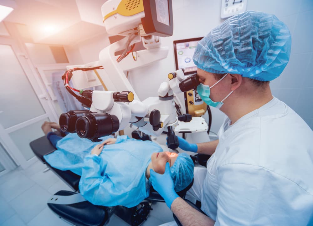
What is MIGS?

The recent most increased interest in glaucoma surgery has been in Minimally Invasive Glaucoma Surgery (MIGS) with formidable results in mild to moderate glaucoma. These are designed to improve the safety of surgical intervention for glaucoma. Although initially coined minimally invasive, the term micro seems more appropriate because it truly differentiates these microscopic ophthalmic procedures from other minimally invasive surgical procedures (i.e., general surgery). Most MIGS procedures enhance physiologic outflow and are aimed at a different patient population than traditional filtration surgery. As opposed to competing with traditional filtering surgery, MIGS seems to be more of an alternative to medical therapy in an effort to address adherence challenges, adverse events, and quality-of-life (QOL) issues with topical medications.

I-stent

The iStent Trabecular Micro-Bypass Stent (Glaukos, Laguna Hills, CA) received FDA approval in 2012. The device is a heparin-coated, non-ferromagnetic titanium stent with a snorkel shape to facilitate implantation. The device is placed using a single-use, sterile inserter through a 1.5mm corneal incision. The iStent itself is the smallest FDA approved device, measuring at 0.3mm in height and 1mm in length. The iStent is a safe minimally invasive glaucoma surgery (MIGS) procedure that can reduce the need for daily use of glaucoma eye drops. A small (1mm) titanium drainage stent is inserted into the eye’s natural drainage channel to lower intraocular pressure. Most clinical trials concluded that iStent implantation with phacoemulsification resulted in a significantly lower, long-term decrease in IOP and number of medications used compared to phacoemulsification alone. No major complications have been reported.

XEN Glaucoma Implant

The XEN Glaucoma Implant (AqueSys Implant) was created by AqueSys Inc acquired by Allergan. The implant itself is made out of a soft, collagen-derived, gelatin that is known to be non-inflammatory. The Xen Gel Stent aims to reduce intraocular pressure by inserting a small drainage tube into the eye. The stent allows fluid to drain from the anterior chamber into a reservoir (bleb) under the conjunctiva. The goal of implantation is to create an aqueous humor outflow path from the anterior chamber to the subconjunctival space. The implant is injected through a small corneal incision with the use of an inserter similar to those used for IOLs. Similar to other implants, it can be performed in conjunction with cataract surgery.
All shunts perform approximately the same function of lowering the eye pressure. It’s important to note that no treatment is guaranteed to completely eliminate the need for eye drops, but the strong results seen in the use of either the Istent or XEN Gel Stent give the right patients a very strong choice for eliminating the need for them.
Your Glaucoma specialist will discuss the best one for you.

How will the shunt affect the appearance of the eye?

I-stent

The I-stent does not appear on the outside of the eye and does not create a bleb outside as it relies on the existing venous outflow system of the eye to help drain the aqueous fluid out.
Initially after surgery, the eye will be red and swollen to a variable degree. The recovery is rapid and redness should be short lived.

XEN Gel Implant:

Successful MIGS surgery takes much less time than many other types of Glaucoma surgery, typically lasting 30 minutes at the most. At Moorfields Eye Hospital Dubai/Abu Dhabi, MIGS implantation is usually performed under local anaesthesia, although general anaesthesia is also possible under certain circumstances.
Typically a drug called Mitomycin C (anti-scarring medication) is also used at the time of surgery if indicated to suppress healing.

The Surgery Itself

Successful MIGS surgery takes much less time than many other types of Glaucoma surgery, typically lasting 30 minutes at the most. At Moorfields Eye Hospital Dubai/Abu Dhabi, MIGS implantation is usually performed under local anaesthesia, although general anaesthesia is also possible under certain circumstances.
Typically a drug called Mitomycin C (anti-scarring medication) is also used at the time of surgery if indicated to suppress healing.

Complications at the time of surgery

Complications occurring at the time of MIGS surgery are extremely rare with an excellent safety profile. Complications when they do occur, tend to occur during the first 2 weeks after the surgery (see Success rates and complications).

After Surgery – Postoperative Care

The day of surgery and the next day

You will usually be discharged home from hospital later the same day after surgery. It is usually necessary for the operated eye to be examined again one day after surgery.
The eye is normally patched the first night after surgery and the patch removed the following day. If the un-operated eye does not see well, then the operated eye will not be patched. Instead, a clear shield will be placed on the operated eye so that you will still be able to see to get around after surgery.

What should I expect to feel during the postoperative period?

It is normal for the vision to be blurred and the eye to be uncomfortable after surgery. The period of blurring is variable. The blurring is usually worst for the first 1 to 2 weeks after surgery, and improving slowly afterwards. It takes about 1 month for the eye to feel completely normal though vision would stabilise much earlier.

Eye Drops

Eye drops and tablets to lower the eye pressure are not normally required for the operated eye during the first night after surgery, unless the surgeon recommends that you continue to use them. It is important to continue any eye drops for the un-operated eye unless advised otherwise.
The following day, the postoperative eye drops are usually started after removal of the eye patch and cleaning of the eye. The postoperative eye drops will usually consist of an antibiotic and anti-inflammatory steroid eye drops to use for the first month after surgery. You will be advised if any changes in these are required at each clinic visit.
To start with the steroid eye drop will be used intensively (every 2 hours or about 8 times daily) during the day and the antibiotic 4 times daily.

Postoperative visits to clinic

As the intraocular pressure may be too high or too low in the first week after shunt surgery, the patient is usually examined in clinic two weekly for approximately the first month, with visits reducing in frequency after that. If indeed the pressure is too high or too low, rest assured that your specialist will manage this appropriately.

Activity and Instructions of care after MIGS

Following surgery you are able to read and watch television as normal as these activities will not harm your eye. It is however important to avoid strenuous activity during the first few weeks after surgery. The following table is a general guide to do’s and don’ts.
IF ANYTHING DO NOT RUB/BUMP/PRESS THE EYE. IN DUSTY ENVIRONMENTS PLEASE WEAR EYE SHIELD OR YOUR OWN GLASSES. DO NOT STOP YOUR EYE DROPS UNLESS THE DOCTOR ADVISES. DO NOT USE ANY OTHER PRODUCTS IN THE EYE.
If in doubt please ask your doctor or nurse in clinic.

When can I go back to work?

The duration of time off work will depend on a number of factors such as the nature of your employment, the state of the vision in the other eye and the pressure in the operated eye.
Typically someone working in an office environment would require 1 week off if the postoperative course is smooth. Someone whose occupation involves heavy manual work or work in a dusty environment may require 2 weeks or upto 1 month (e.g. builders, working in dessert).

When is the eye back to normal?

It takes 1 month for the eye to feel completely normal in most cases, and sometimes longer in more complicated cases. At that point, the patient will usually have a refraction (spectacle) test as often the spectacle prescription will be slightly different than before surgery.

Success Rates and Complications

Success rates

Most glaucoma surgical studies examine success rates over a 5 year period. The I-stent and XEN gel implant are showing good results with 80-90% qualified success in most studies. Although a sizable proportion of patients achieve good pressure control without the need for continued glaucoma medication, many patients still require some medication to assist the shunt in controlling the pressure especially if they are on more than one drop. In such circumstances, the medication required is usually less than that required before the surgery.

Complications

As with any surgery, there is a potential for complication or problems to arise. Complications can occur during the surgery, shortly after the surgery or many months after surgery.
Severe complications are rare and may happen either if the eye pressure drops very low, or very quickly during the early postoperative period, or if the eye becomes infected.
Bleeding
The most serious problem that can occur is bleeding inside the eye. This can lead to loss of vision and even blindness but occurs in less than 1 in 1000 patients. There are measures taken to prevent this but this is not a predictable complication.
Infection
An infection inside the eye can be very serious and also cause loss of vision or blindness. This also happens in less than 1 in 1000 patients. Measures are taken and aseptic techniques used to prevent this complication.
Eye Pressure
After the operation the eye pressure may be too high or too low. This may require additional treatment in the outpatient clinic or sometimes further surgery is required.
Inflammation
This can occur inside the eye and is usually treated with eyedrops.
If the operation is not performed in a timely manner then there is potential for further irreversible visual loss due to high pressure/fluctuating pressure in the eye leading to blindness.
However the informed decision rests with the patient and your Glaucoma specialist will be available and very happy to discuss your concerns and expectations before proceeding with surgery.

Disclaimer

Accuracy

While every step has been taken to compile accurate information and to keep it up to date, we cannot guarantee its correctness and completeness. The information provided in this information sheet is designed as an adjunct to, and not a substitute for professional healthcare advice, by a qualified doctor or other healthcare professional, which will be tailored to a patient’s individual circumstances. Dr Sohaib Mustafa and Moorfields Eye Hospital Dubai/Abu Dhabi cannot take responsibility if you rely solely on the information in this information sheet.