Age-related Macular Degeneration (AMD)

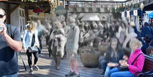
The central part of the retina (at the back of the eye) is called the macula and it has an important function as it controls the quality and sharpness of the central part of our vision.
Over time, the macular can wear out and lose some of its functionality which means loss of central vision (not the peripheral vision) and this can cause problems, when it comes to everyday tasks such as reading
The good news is that the deterioration of vision is usually happens quite slowly.
However, there are two types of macular degeneration – ‘wet’ and ‘dry’ – and what is known as the ‘wet’ form results in a sudden loss of central vision, which is a medical emergency and urgent treatment is needed.

Macula

The macula is a small, extremely important area at the centre of the retina, the light-sensing tissue at the back of the eye and is responsible for seeing fine details clearly.

With AMD (Age-related Macular Degeneration), you lose the ability to see fine details, close-up and at a distance. 

AMD (Age-related Macular Degeneration)affects only your central vision and side and peripheral vision usually remains normal.  For example, when people with AMD gradually lose the ability to recognise people’s faces. 

Types of AMD (Age-related Macular Degeneration)

There are two types of AMD (Age-related Macular Degeneration).
Most people (about 75%) have a form called “early” or “dry” AMD, which develops when there is a build-up of waste material under the macula and thinning of the retina at the macula.  Most people with this condition have near normal vision or milder sight loss in the initial stage.
10% of patients with dry AMD can progress to wet AMD. Wet AMD occurs when abnormal blood vessels grow underneath the retina and leak blood and fluid, which can prevent the retina from working properly.
Eventually the bleeding and scarring can lead to severe permanent loss of central vision, but the eye is not usually at risk of losing all vision (going ‘blind’) as peripheral vision remains. Symptoms of wet AMD include sudden central blurriness and distortion, where straight lines appear as wavy (metamorphopsia).
A late stage AMD is called geographic atrophy, where vision is lost through severe thinning or even loss of the macula tissue without any leaking blood vessels.

Treatments for AMD

Intravitreal injections (injections into the eye) for wet AMD use a medicine called anti-VEGF of which there are 2 licensed versions Eylea and Lucentis.

When anti-VEGF medicines are injected into the eye on a regular basis, it can stop the abnormal blood vessels growing, leaking and bleeding under the retina. 

Most people with wet AMD need to have these injections several times a year, so patients need to get their care closer to home.  Laser treatment is also available for specific forms of AMD, but is not effective for most cases. 

There is currently no treatment for dry AMD.

Retinal Detachment

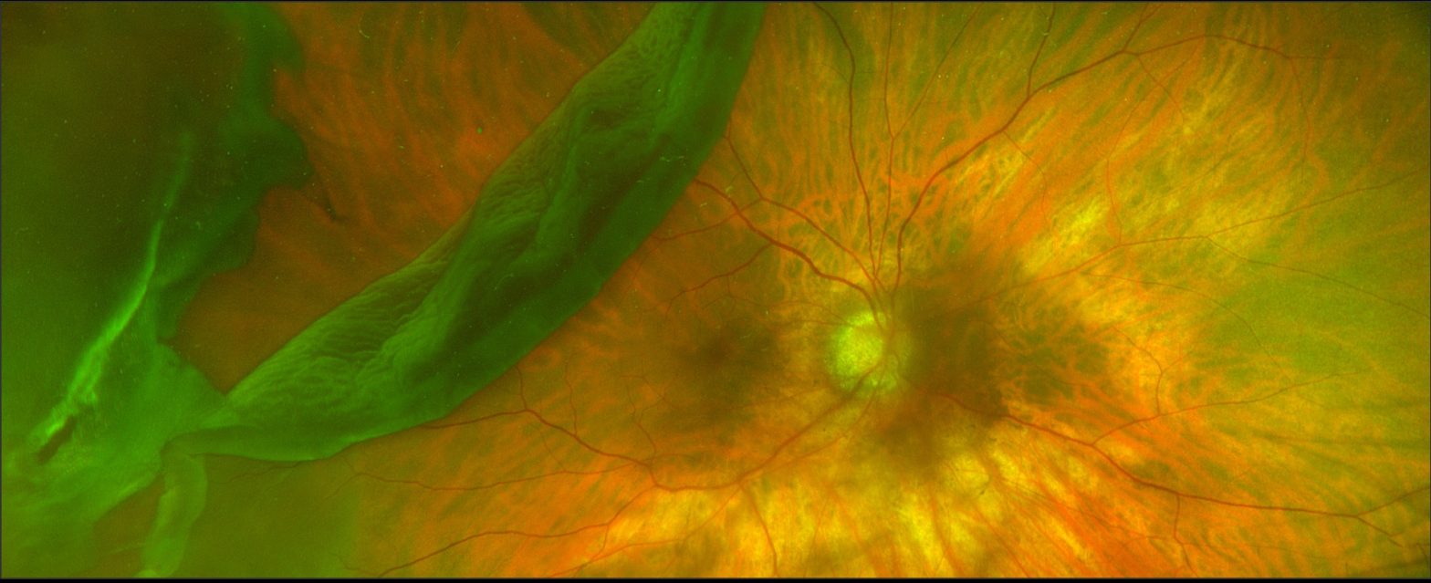
Retinal detachment is a condition when the thin lining at the back of the eye (the retina) begins to come away and separate itself from the underlying wall of the eye (the choroid) which contains blood vessels that supply it with vital oxygen and nutrients.
If not treated promptly, retinal detachment will lead to blindness in the affected eye.
A retinal detachment is usually caused by a tear in the retina and this is termed a Rhegmatogenous retinal detachment. There are other types of retinal detachment namely Traction retinal detachment which is usually seen in advanced diabetic retinopathy and Exudative retinal detachment (usually seen in people with inflammation). It is the Rhegmatogenous retinal detachment which needs urgent surgery in most cases.

Symptoms

Most people experience advance symptoms of a possible retinal detachment before losing their sight, including:

  • sudden appearance of floaters – black dots, specks or streaks that float across the field of vision (usually only one eye is affected)
  • web effect of lots of small floaters – some people see a single large black floater in the shape of a fly
  • sudden short flashes (no more than one second) of light in the affected eye
  • seeing a dark shadow like a curtain falling down from the outer aspect of the field of vision in the affected eye
  • blurring or distortion of vision

Without immediate treatment, the vision in the affected eye will start to deteriorate, creating the effect of a shadow or ‘black curtain’ spreading across the vision. If a person experiences a shadow in the vision then It is extremely important to consult an eye doctor immediately before the shadow spreads across the centre of the vision.
Retinal detachment usually only occurs in one eye, however if one eye is affected, there is a 1 in 10 chance that the retina in your other eye will also detach.
You should contact your eye doctor immediately if you get any of these warning signs.

Causes

The retina at the back of the eye sends signals to the brain, enabling us to see. Without a blood supply, the retina’s nerve cells die leading to loss of sight.
Retinal detachment is usually the result of the retina becoming thinner and weaker with age. This causes a tear in the retina and once a tear develops the retina can pull away from the underlying blood vessel wall (the choroid).
Very short-sighted people have the greatest risk of developing age-related retinal detachment (though the risk is still very small) because they are often born with a thinner than normal retina in the first place.
Previous eye surgery, such as cataract removal, may also make the retina more vulnerable to damage.
In some cases, a tear can develop if the eye is suddenly injured, such as by a blow to the face but this is less common.

Diagnosis

If the eye specialist (ophthalmologist) suspects a retinal detachment, an examination of the back of the eye will confirm it.

Retinal Detachment Treatment

Urgent Retinal Detachment treatment is essential to lower the risk of permanent vision loss. The Retinal Detachment treatment is mainly surgery and eye drops will not help.
Most but not all detached retinas can be successfully reattached through different types of surgery, followed by a few months of recovery when vision will be weak, affecting lifestyle activities such as driving.
Some people’s eyesight does not fully return after surgery and they have permanently reduced peripheral (side) or central vision. This can happen even if the retina is reattached successfully.
This risk is higher the longer the detachment was left untreated.

Who does it affect?

Retinal detachment is rare. For example, only one in every 10,000 people will develop it in any given year in the UK.
As retinal detachment is associated with ageing, most cases affect older adults aged between 60 and 70. Retinal detachment caused by an injury can affect people of any age, including children.
Contact Moorfields Dubai for Retinal Detachment treatment today.

Moorfields Eye Hospital Dubai treats cataract and extreme short sightedness with the most powerful custom-made lenses ever implanted in a patient in the UAE

[:en]robert
20 September 2016 (Dubai – United Arab Emirates): A team from Moorfields Eye Hospital Dubai has successfully treated a patient who required a complex surgical implant of extremely powerful intraocular lenses (IOL) to treat her cataract (clouding of the eye’s natural lens) and correct her high myopia (extreme short sightedness). As a result of implanting two lenses with the highest minus power ever implanted in the UAE, the patient’s vision and quality of life have improved dramatically. Standard IOLs were not an option and the lenses were delivered from Germany, custom-made specifically for the patient.
The patient, who is a UAE resident, had a complicated history of eye problems including cataract and was unable to see without very thick lenses, due to her extreme short sightedness. The complexity of the treatment included the challenge of very accurately calculating the power of the lenses required to correct the patient’s vision, manufacturing the custom lenses, and finally the surgical procedure to implant correctly such powerful lenses.
Professor Robert Scott, Medical Director at Moorfields Eye Hospital Dubai, comments: “Though complex, the bilateral surgery to implant these special intraocular lenses was performed without complications and the results have been excellent. Our patient presented us with the challenge of treating her cataract and correcting her extreme short sightedness, which meant that we had to arrange the custom-made lenses with the most minus refractive reading ever implanted in the UAE. We are all delighted that she is now able to see very well and only needs her light glasses for reading; we believe the surgery to improve her vision will significantly enhance her life.”
According to the patient, opening her eyes for the first time after the surgery was “an amazing experience and the world seemed brighter and clearer without contact lenses and like seeing the world in a new light. It feels like I’ve started a new chapter in my life. I’m very pleased that I had the surgery done at Moorfields Eye Hospital Dubai where the expert team gave me the confidence and support needed for successful surgery. I can’t thank Professor Robert Scott, the anesthesiologist and nurses at Moorfields enough for their kindness and expertise.”
Cataract refers to the clouding of the natural lens in the eye; it is the most cause of vision loss in people over the age of 40 and is the leading cause of blindness in the world. Myopia is a very common condition that affects around 25% of the general population. High myopia (extreme short sightedness) is very difficult to treat/correct using standard contact lenses or spectacles, because of the thickness of the lenses needed to correct the vision. This is where an intraocular lens (IOL) – a synthetic artificial lens implanted into the eye to replace the natural lens – is a very effective treatment.

Glaucoma Surgery Treatment

What is Glaucoma?

Glaucoma is a condition in which the fluid which is produced by the eye – known as the aqueous humour – does not drain away normally and this results in increased pressure inside the eye. If this condition is not treated, it can lead to damage of the optic nerve which connects the eye to the brain and loss of vision.

Signs and Symptoms

Glaucoma does not present symptoms until late stages, when it can cause damage to your field of vision. It generally starts at the periphery of the field of vision and then leads to a gradual narrowing or tunneling of the vision. If treatment is delayed, these defects can severely affect your vision and can cause “tunnel vision”.

Importance of Early Diagnosis

Early treatment can prevent damage from occurring, however, any damage that occurs is permanent. Therefore it is very important to diagnose the condition early to maintain the maximum level of peripheral vision. Risk factors for Glaucoma include short sightedness, diabetes and people with a family history.
If there is a history of glaucoma in your immediate family then regular eye checks are essential. Ideally, everyone over the age of 40 should have an eye test for glaucoma.

Tests for Glaucoma

At Moorfields Eye Hospital Dubai we have the latest equipment for diagnosing and staging glaucoma.

Pressure Check

A local anaesthetic drop which lasts only for 10-20 minutes will be placed in both eyes, then a small instrument is placed gently against the eye. This will measure the pressure inside the eye. Alternatively the pressure may be checked using air. A small puff of air is blown into your eye, this is not painful and gives a quick and accurate measurement of the pressure.

Pachymetry

This measures the thickness of the cornea by means of an ultrasound probe. It has proven to be important in determining precisely the Intraocular pressure.

Treatment

The treatment for Glaucoma focuses on reducing the pressure inside the eye, by lowering the amount of fluid being produced or by improving the drainage of the fluid away from the eye.
The usual treatment
is with eye drops that best suit your case. If you are prescribed eye drops, they must be taken regularly and according to the prescribed doses.
In cases resistant to medical treatment, surgery may need to be performed.
Ask our staff for glaucoma screening and follow-up packages which conveniently include all the above tests.
General Examination
The examination includes looking directly at the optic nerve at the back of the eye for any signs of damage.
Visual Field Testing
The peripheral vision is tested to see whether there has been any loss of vision at the outer edge of the field of vision.
Optical Coherence
Detecting Glaucoma early, before symptoms are apparent, is important and Tomography helps achieve this and so reduces the risk of any damage to the optic nerve.