عن الحالة
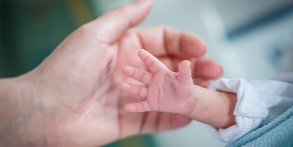
اعتلال الشبكية لدى الخدّج هي مشكلة في العيون تؤثر على المواليد الخدّج. ويصاب الطفل بهذه المشكلة عندما لا يكتمل تطور الشبكية لديه في الأسابيع اللاحقة لولادته. نتيجة للولادة المبكرة، فقد تتشكل أوعية دموية غير طبيعية تتصف بهشاشة جدرانها وقابليتها للتسريب، كما تؤدي إلى إحداث ندوب في الشبكية ونزعها من مكانها. تؤدي هذه الحالة إلى انفصال الشبكية الذي يعتبر السبب الرئيسي في اعتلال الرؤية والعمى المرتبط بها.
بعض حالات الإصابة باعتلال الشبكية لدى الخدّج طفيفة ولا تحتاج إلى أي علاج. ولكن هناك حالات شديدة تستدعي التدخل الجراحي لمنع فقدان البصر. وهناك الجراحة بواسطة الليزر لمنع نمو الأوعية الدموية المختلة وضمان ألا يحدث انفصال الشبكية.
ليس لاعتلال الشبكية لدى الأطفال الخدّج علامات أو أعراض، والطريقة الوحيدة للتحقق من الحالة هي فحص أخصائي طب عيون مؤهل لعيون حديثي الولادة.
تصنيف اعتلال الشبكية لدى الأطفال الخدّج
يصنف اعتلال الشبكية لدى الأطفال الخدّج وفق مدى خطورته، إلى
- مراحل (0 إلى 5)
- موقعه في 3 مناطق (1 – 3)
- مدى المرض وفق الساعات (1 – 12)
- وجود مرض آخر.
عوامل المخاطر
الوزن عند الولادة ومدة الحمل
الفئة الأكثر عرضة للخطر هم المواليد ذوي الوزن الخفيف للغاية ويحتاجون إلى العلاج سريعاً. وبالمثل، هناك علاقة عكسية بين مدة الحمل وتزايد احتمال الإصابة بالمرض.
استعمال الأكسجين
برغم عدم التثبت علمياً من العلاقة الدقيقة بين العلاج بالأكسجين وهذه الحالة، إلا أن من الواضح أن للعلاج بالأكسجين دور مهم في التسبب فيها. قد تؤدي الإدارة الصارمة لإيصال الأكسجين من دون تغيير وتفاوت ورصد ذلك إلى تراجع الإصابة باعتلال الشبكية لدى الأطفال الخدّج.
الفحص
يجب فحص جميع الأطفال الذين تقل أعمار حملهم عن 32 أسبوعًا (حتى 31 أسبوعًا و6 أيام) أو من يزنون أقل من 1501 جرامًا عند الولادة للتأكد من عدم إصابتهم باعتلال الشبكية. يعتمد الوقت الدقيق الذي يتم فحصه فيه على عمر الحمل للطفل و/أو الوزن عند الولادة. تعتمد الفترة الزمنية بين الفحوصات على نتائج الفحص الأول. يجري مناقشة ذلك مع طبيب عيون الأطفال.
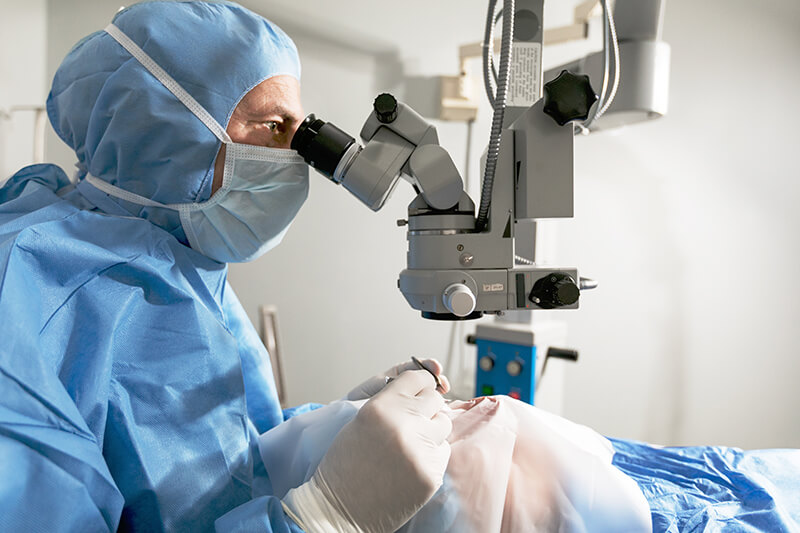
يشمل الفحص توسيع الحدقة (الجزء الأسود من العين) بقطرات العين للسماح برؤية كاملة للشبكية (الجزء الخلفي من العين). عقب ذلك، يفحص الطبيب عيني الطفل بأدوات تكبير مناسبة؛ وقد يلتقط صورًا للجزء الخلفي من العين.
يمكن إيقاف الفحص عند التأكد من استبعاد إصابة الطفل باعتلال الشبكية.
الهدف من الجراحة هو إيقاف نمو الأوعية الدموية غير الطبيعية من خلال تركيز العلاج على الشبكية الطرفية (جوانب الشبكية) للحفاظ على الشبكية المركزية (أهم جزء في الشبكية). تركز الجراحة على مناطق الندبات في الشبكية الطرفية لوقف النمو غير الطبيعي ومنع شد الشبكية.
يتوقف تحديد علاج اعتلال شبكية الأطفال الخدج على شدته (مرحلته، منطقته، ووجود أمراض أخرى)، وكذلك مدى تقدم الحالة ووزن الطفل عند ولادته وعمر الحمل والعرق الذي ينتمي إليه الطفل.
التوقيت
عند التشخيص، يتوجب المبادرة بالعلاج، خلال يومين إلى 3 أيام من التشخيص. وذلك لأن هذا المرض يتفاقم بسرعة كبيرة وقد يسبب الضرر البالغ لعين الطفل وقدرته على الإبصار في حال عدم علاجه بدقة وسرعة.
التخدير
تُجرى جراحة اعتلال الشبكية للطفل الخديج إما تحت التخدير العام (من خلال دواء يحافظ على المريض في حالة شبيهة بالنوم العميق) أو التخدير العميق (من خلال دواء يجعله غير مدرك لخطوات العملية ولكنه ليس مخدرًا بعمق مثل التخدير الكلي).
أنواع العلاج
العلاج بالليزر
يستخدم علاج التخثير الضوئي بالليزر لحرق المناطق المحيطة بحافة الشبكية. هذا هو العلاج المفضل، لكونه الأقل حدة والأدنى ضرراً لعين الطفل. كما أن الليزر أسهل في تطبيقه في علاج حالات مؤخرة الشبكية.
مضاعفات العلاج بالليزر:
- حروق في القرنية والقزحية
- الساد
- نزف في الشبكية والجسم الزجاجي
العلاج بالتجميد
تستخدم المعالجة بالتجميد أداة لتجميد جزء معين من العين يمتد متجاوزًا حواف الشبكية. وهو يأتي في المرتبة الثانية بعد العلاج بالليزر لأن مضاعفاته أكثر.
مضاعفات العلاج بالتجميد:
- وذمة الجفن
- جرح الملتحمة
- نزف في مقدمة الشبكية والجسم الزجاجي
- مضاعفات من قبيل بطء نبضات القلب والزرقة والاكتئاب التنفسي
أدوية العامل المضاد لنمو بطانة الأوعية الدموية (anti-VEGF)
تعمل عقاقير العامل المضاد لنمو بطانة الأوعية الدموية على منع فرط نمو الأوعية الدموية في الشبكية.
مضاعفات أدوية العامل المضاد لنمو بطانة الأوعية الدموية
- انفصال الشبكية
- الساد
- التهاب باطن المقلة
- تقلص التكاثر الزجاجي المصاحب لاعتلال الشبكية
جراحة الجسم الزجاجي
في المرحلة المتقدمة من اعتلال الشبكية، يشمل العلاج:
بعج الصلبية – يمكن استخدامه، ويتضمن وضع شريط مرن مصنوع من السيليكون حول محيط العين. يوضع الشريط حول الصلبة، أو أبيض العين، ليدفعها أو “يبعجها”. وهذا بدوره يدفع الشبكية الممزقة إلى الاقتراب من الجدار الخارجي للعين والبقاء فيه.
استئصال الجسم الزجاجي، وهي جراحة معقدة تنطوي على استبدال الجسم الزجاجي، أو الجل الشفاف في وسط العين، بمحلول ملحي. تسمح عملية إزالة الجسم الزجاجي بإزالة أنسجة الندوب وتخفف من سحب الشبكية. وهو ما يمنع الشبكية من الانفصال.
ما بعد العملية
إذا لم يكن الدخول إلى المستشفى ضروريًا، فستتمكن من اصطحاب طفلك إلى المنزل بعد حوالي ساعة من العملية. تتضمن رعاية المتابعة للجراحة وضع قطرات في عين طفلك (لمنع العدوى) لمدة أسبوع على الأقل.
ضماناً لشفاء العينين بشكل سليم وعدم وجود اعتلال في الشبكية، يجب جدولة فحوصات العين بناءً على تعليمات طبيب العيون. ويكون ذلك في العادة كل أسبوع أو أسبوعين.
المخاطر والمضاعفات
الهدف من جراحة اعتلال الشبكية لدى الخدج هو منع تدهور الحالة والعمى. على الرغم من أن للجراحة معدل نجاح جيد، إلا أن ليس جميع الأطفال يستجيبون للعلاج. قد يفقد ما يصل إلى 25٪ من الأطفال الذين خضعوا للجراحة بعض أو كل قدرتهم على الإبصار.
في بعض الحالات، لا تنجح الجراحة بالليزر أو العلاج بالتجميد (مما يعني أن الأوعية الدموية غير الطبيعية تستمر في النمو) وقد يلزم تكرار العلاج. إذا استمر النمو، فقد يكون من الضروري محاولة بعج الصلبة أو استئصال الجسم الزجاجي. ومع ذلك، من المهم أن تعرف أن الجراحة للمراحل المتقدمة من اعتلال الشبكية لدى الخدج لا تضمن إعادة شبكية العين لحالتها وإعادة البصر بقدرته المعتادة.
ضع في اعتبارك أنه بالنسبة لجميع أنواع جراحة اعتلال الشبكية للخدج سوف يفقد الطفل درجة من درجات الرؤية المحيطية (الجانبية). وحتى إذا توقف تدهور اعتلال الشبكية، فلا يزال من الممكن أن تتأثر الرؤية بالطرق التالية:
- الحول: خلل في انتظام العينين
- الغمَش (العين الكسولة): عين أضعف من الأخرى
- حسر البصر: قصر النظر أي صعوبة في رؤية الأشياء البعيدة بوضوح
- الزرَق: ضغط متزايد على العين بما يمكن أن يتلف العصب البصري ويتسبب في فقدان البصر.
- انفصال الشبكية: انفصالها عن العين بما يتسبب في العمى.
وفي ظل إمكانية حدوث فقدان البصر ومضاعفات، يجب أن يخضع أي طفل خاض جراحة اعتلال الشبكية لفحوصات سنوية ومنتظمة حتى سن البلوغ.
اخصائيين اعتلال الشبكية لدى الأطفال الخدّج
د. عمران جاويد
استشاري طب العيونمتخصص في طب عيون الأطفال و جراحة الحول
د. محمد عرفان خان
استشاري طب العيون في طب عيون الأطفال وجراحة الحول عند البالغين
د. سيد علي
استشاري طب العيون للأطفال تخصص في الحول والكتراكت
د. محمد عرفان خان
استشاري طب العيون في طب عيون الأطفال وجراحة الحول عند البالغيناطلب موعد
